. 2020 May;71(5):1750-1765.
doi: 10.1002/hep.30929. Epub 2019 Dec 30.

Monoacylglycerol Lipase Inhibition Protects From Liver Injury in Mouse Models of Sclerosing Cholangitis

Affiliations

Monoacylglycerol Lipase Inhibition Protects From Liver Injury in Mouse Models of Sclerosing Cholangitis

Matteo Tardelli et al. Hepatology. 2020 May.

Abstract

Background and aims: Monoacylglycerol lipase (MGL) is the last enzymatic step in triglyceride degradation, hydrolyzing monoglycerides into glycerol and fatty acids (FAs) and converting 2-arachidonoylglycerol into arachidonic acid, thus providing ligands for nuclear receptors as key regulators of hepatic bile acid (BA)/lipid metabolism and inflammation. We aimed to explore the role of MGL in the development of cholestatic liver and bile duct injury in mouse models of sclerosing cholangitis, a disease so far lacking effective pharmacological therapy.

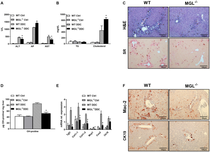
Approach and results: To this aim we analyzed the effects of 3,5-diethoxycarbonyl-1,4-dihydrocollidine (DDC) feeding to induce sclerosing cholangitis in wild-type (WT) and knockout (MGL-/- ) mice and tested pharmacological inhibition with JZL184 in the multidrug resistance protein 2 knockout (Mdr2-/- ) mouse model of sclerosing cholangitis. Cholestatic liver injury and fibrosis were assessed by serum biochemistry, liver histology, gene expression, and western blot characterization of BA and FA synthesis/transport. Moreover, intestinal FAs and fecal microbiome were analyzed. Transfection and silencing were performed in Caco2 cells. MGL-/- mice were protected from DDC-induced biliary fibrosis and inflammation with reduced serum liver enzymes and increased FA/BA metabolism and β-oxidation. Notably, pharmacological (JZL184) inhibition of MGL ameliorated cholestatic injury in DDC-fed WT mice and protected Mdr2-/- mice from spontaneous liver injury, with improved liver enzymes, inflammation, and biliary fibrosis. In vitro experiments confirmed that silencing of MGL decreases prostaglandin E2 accumulation in the intestine and up-regulates peroxisome proliferator-activated receptors alpha and gamma activity, thus reducing inflammation.

Conclusions: Collectively, our study unravels MGL as a metabolic target, demonstrating that MGL inhibition may be considered as potential therapy for sclerosing cholangitis.


Figures

Figure 1
Figure 1
MGL−/− mice display reduced biliary fibrosis and inflammation after 2 weeks of DDC feeding. Cholestatic liver injury resembling PSC was induced in C57BL/6J and MGL−/− mice (n = 8 per group) by 2 weeks feeding with DDC. (A) Serum biochemistry reflects improved levels of transaminases (ALT and AST but unchanged AP) as well as (B) increased cholesterol levels and unchanged TG. (C) Representative H&E images (×10 magnification) with markedly improved liver histology and ameliorated fibrosis in line with (D) reduced hepatic hydroxy (OH)‐proline levels in MGL−/− mice fed DDC. (E) Hepatic gene expression of profibrogenic markers Tgfβ, Col1α1, and Col1α2 diminished while proinflammatory cytokines/chemokines Mcp1, Cox2, and Ck19 were reduced after DDC feeding. (F) Representative images of Mac‐2 and CK19 staining in liver tissue. White/gray bars represent mice fed the control diet; black and dashed bars represent mice fed the DDC diet. Results are expressed as mean ± SD. *P < 0.05 for MGL−/− DDC versus WT DDC mice. Abbreviations: Ctrl, control; SR, sirius red.
Figure 2
Figure 2
MGL deletion down‐regulates cholesterol synthesis while increasing FA synthesis and oxidation. (A) Hepatic gene expression of Srebp2, Hmgcr, and Ldlr indicates diminished cholesterol synthesis. (B) Hepatic expression of FA synthesis genes increased as seen for Srebp1c, Pparγ2, and Cd36 gene. (C) Mitochondrial β‐oxidation increased as evidenced by Pparα, Cpt1α, Pgc1α, Aox, and (D) Hmox1 and Nrf2 gene expression. In line with (E) western blot analysis of HO‐1 (calnexin shown as loading control). (F) Hepatic ATP content measured in cytosol and mitochondria from liver homogenates increased in MGL−/− DDC. Results are expressed as mean ± SD. *P < 0.05 for MGL−/− DDC versus WT DDC mice (n = 8). Abbreviation: Ctrl, control.
Figure 3
Figure 3
BA synthesis and export are increased, whereas import is unchanged in MGL−/− mice fed DDC. (A) Hepatic gene expression of limiting BA synthesis pathway (Cyp7a1) and detoxification (Cyp3a11, Cyp2b10). (B) Gene expression profiling of Ntcp, Oatp1, Mrp2, Mrp3, Mrp4, Bsep. (C) Representative western blot with (D) corresponding densitometry (n = 2 per group) of BA transporters showing augmented Mrp2, unchanged secretion (Bsep), and unchanged uptake (Oatp1 and Ntcp), Calnexin was used as a loading control. Results are expressed as mean ± SD. *P < 0.05 for MGL−/− DDC versus WT DDC mice (n = 8). Abbreviation: Ctrl, control.
Figure 4
Figure 4
Intestinal inflammation diminishes in MGL−/− mice despite linoleic acid and AA accumulation. (A) Intestinal gene expression of proinflammatory genes Tnfα, F4/80, and Cox2 decreased in MGL−/− mice, whereas Pparγ, Pparα, and Cpt1α increased despite Fgf15 down‐regulation. (B) Gas chromatography quantification of intestinal polyunsaturated FAs evidenced accumulation of 18:2w6 (linoleic acid) and 20:4w6 (AA) in MGL−/− mice fed DDC (n = 4). (C) PGE2 levels decreased in intestine and liver (trend wise). (D) Intestinal gene expression of alternative lipid‐hydrolyzing enzymes Abhd6 and Abhd12 were up‐regulated in MGL−/− mice fed DDC. (E) DDC treated MGL−/− mice showed significantly different microbiome from WT DDC (dot plot) with reduced abundance of Proteobacteria (bar graph). Results are expressed as mean ± SD. *P < 0.05 for MGL−/− DDC versus WT DDC mice (n = 8). Abbreviation: Ctrl, control.
Figure 5
Figure 5
The MGL inhibitor JZL184 ameliorates biliary fibrosis and inflammation in Mdr2−/− mice. Eight‐week‐old Mdr2−/− mice were fed JZL184 for 4 weeks (n = 9 per group). (A) Serum biochemistry evidenced decreased serum levels of ALT and AP, with unchanged AST and (B) diminished plasma BA in JZL184‐fed mice but not in Mdr2−/− mice receiving chow. (C) Representative H&E images (×10 magnification) with markedly improved liver histology and ameliorated fibrosis (sirius red) confirmed by (D) diminished hepatic OH‐proline levels. (E) Hepatic gene expression of Col1α1, Cyp7a1, Ck19, and Vcam‐1 and proinflammatory cytokine Mcp1 diminished. (F) Representative Mac‐2 and CK19 IHC pictures show reduced cholangiocyte proliferation and inflammation in JZL184‐treated Mdr2−/− mice. Results are expressed as mean ± SD. *P < 0.05 for Mdr2−/− in white bars versus Mdr2−/− mice fed JZL184 in black bars and WT gray bars. Abbreviation: SR, sirius red.
Figure 6
Figure 6
JZL184 feeding increases hepatic β‐oxidation, also leading to diminished inflammation in the intestine. (A) Gene expression of Pparγ1, Pparγ2, and Cd36 and (B) Pparα, Cpt1α, Pgc1α (in trend), Hmox1, and Nrf2 (in trend) is up‐regulated. (C) Hepatic ATP content increased in mitochondria but not in cytosol. (D) Intestinal gene expression of Pparα and Cpt1α is up‐regulated, whereas Pparγ2 and Fgf15 remained unchanged; inflammatory markers F4/80 and Cox2 are strongly decreased. (E) Gas chromatography quantification of polyunsaturated FAs in the intestine from Mdr2 −/− mice fed JZL184 evidenced enrichment of AA. (F) Microbiome analysis showed changed microbial composition in Mdr2 −/− mice fed JZL184, with reduced abundance of Ruminococcaceae compared to Mdr2 WT. Results are expressed as mean ± SD. *P < 0.05 for Mdr2−/− versus Mdr2−/− JZL184‐fed mice (n = 9). Abbreviation: NMDS, nonmetric multidimensional scaling.
Figure 7
Figure 7
The MGL inhibitor JZL184 mitigates cholestatic injury after DDC feeding. WT mice received 2 weeks of DDC feeding and 4 days of JZL184 to investigate disease resolution. (A) Serum biochemistry reflects diminished levels of serum transaminases (ALT and trend for AST but unchanged AP) and (B) bilirubin. (C) Representative H&E images (×10 magnification) with improved liver histology and ameliorated fibrosis (sirius red) in WT animals fed DDC+JZL184. (D) Hepatic gene expression of profibrogenic markers Tgfβ and Col1α1 diminished, while proinflammatory cytokines/chemokines Mcp1 and Cox2 remained unchanged and Ck19 diminished. (E) Hepatic gene expression of Srebp1c and Srebp2 was unchanged, Fasn diminished, whereas Aox and Pparα increased. (F) Representative images of Mac‐2 and CK19 staining in liver tissue showing ameliorated inflammation and cholangiocyte reactive phenotype. Gray bars represent control mice fed chow; white bars represent DDC + chow diet; black bars represent mice fed the DDC + JZL184 diet. Results are expressed as mean ± SD. *P < 0.05 for WT DDC + chow versus DDC + JZL184. Abbreviations: Ctrl, control; SR, sirius red.
Figure 8
Figure 8
Silencing MGL in intestinal cells up‐regulates PPAR‐α and PPAR‐γ in vitro, and AA binds PPARs/RXR, diminishing inflammation. MGL was silenced in Caco2 cells. (A) Representative western blot with corresponding Image J quantification showing successful silencing at 5 µM concentration. (B) Caco2 siMGL had induction of Pparγ, Pparα, and Cpt1α with unchanged Fgf19 gene expression and decreased Fxr. (C) Gene expression of proinflammatory genes Tnfα and Cox2 down‐regulated after treatment with AA in siMGL Caco2 cells. (D) Transfection of PPARα, PPARγ, and RXR showed increased luciferase activity of PPRE after AA incubation. (E) Cotransfection of RXR/FXR shows that AA antagonizes induction of FXRE by CDCA. Results are expressed as mean ± SD of three independent experiments in duplicate, *P < 0.05 for siMGL versus siMGL+CDCA and AA treatment versus control or CDCA+AA treatment versus CDCA alone. (F) MGL deletion ameliorates cholestatic liver disease induced by DDC challenge diminishing fibrosis, inflammation, and FA metabolism/oxidation in the liver. It also induces BA synthesis and detoxification as shown by Cyp7a1/Cyp3a11 induction and BA transport (Mrp4) with unchanged Bsep and increased Mrp2. In the intestine, accumulation of AA binds NRs PPAR‐α, PPAR‐γ, and RXRα, resulting in anti‐inflammatory effects as further demonstrated by diminished PGE2 content. Abbreviations: Chol, cholesterol; Ctrl, control; βox, β‐oxidase.

Similar articles

Cited by

References

    1. Eaton JE, Talwalkar JA, Lazaridis KN, Gores GJ. Lindor KD. Pathogenesis of primary sclerosing cholangitis and advances in diagnosis and management. Gastroenterology 2013;145:521‐536. - PMC - PubMed
    1. Dyson JK, Hirschfield GM, Adams DH, Beuers U, Mann DA, Lindor KD, et al. Novel therapeutic targets in primary biliary cirrhosis. Nat Rev Gastroenterol Hepatol 2015;12:147‐158. - PubMed
    1. Hirschfield GM, Heathcote EJ, Gershwin ME. Pathogenesis of cholestatic liver disease and therapeutic approaches. Gastroenterology 2010;139:1481‐1496. - PubMed
    1. Zechner R, Zimmermann R, Eichmann TO, Kohlwein SD, Haemmerle G, Lass A, et al. FAT SIGNALS—lipases and lipolysis in lipid metabolism and signaling. Cell Metab 2012;15:279‐291. - PMC - PubMed
    1. Poursharifi P, Madiraju SRM, Prentki M. Monoacylglycerol signalling and ABHD6 in health and disease. Diabetes Obes Metab 2017;19:76‐89. - PubMed

Publication types

MeSH terms