. 2020 Feb;62(2):204-216.
doi: 10.1165/rcmb.2019-0245OC.

Cross-Talk between Transforming Growth Factor-β and Periostin Can Be Targeted for Pulmonary Fibrosis

Affiliations

Cross-Talk between Transforming Growth Factor-β and Periostin Can Be Targeted for Pulmonary Fibrosis

Yasuhiro Nanri et al. Am J Respir Cell Mol Biol. 2020 Feb.

Abstract

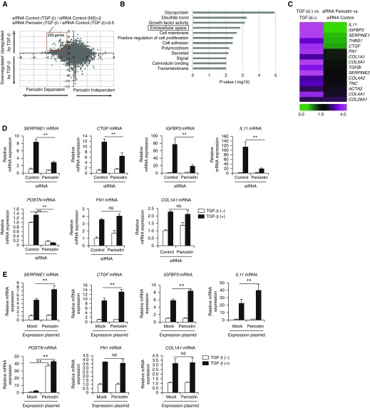
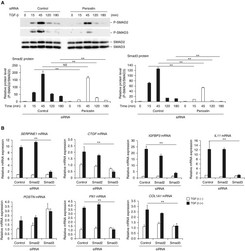
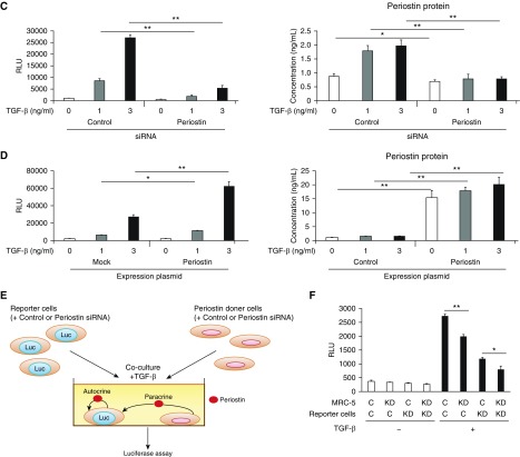
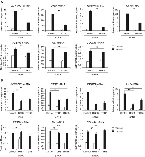
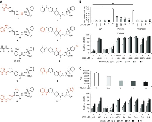
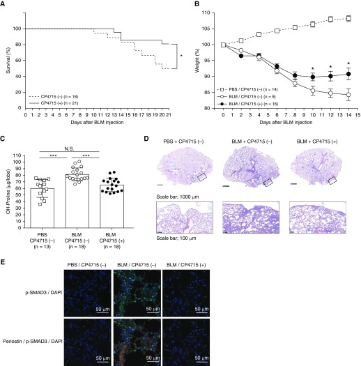
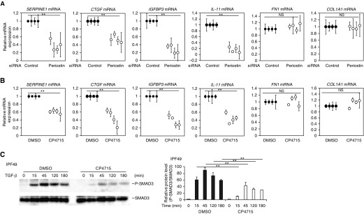
Idiopathic pulmonary fibrosis (IPF) is a devastating disease characterized as progressive and irreversible fibrosis in the interstitium of lung tissues. There is still an unmet need to develop a novel therapeutic drug for IPF. We have previously demonstrated that periostin, a matricellular protein, plays an important role in the pathogenesis of pulmonary fibrosis. However, the underlying mechanism of how periostin causes pulmonary fibrosis remains unclear. In this study, we sought to learn whether the cross-talk between TGF-β (transforming growth factor-β), a central mediator in pulmonary fibrosis, and periostin in lung fibroblasts leads to generation of pulmonary fibrosis and whether inhibitors for integrin αVβ3, a periostin receptor, can block pulmonary fibrosis in model mice and the TGF-β signals in fibroblasts from patients with IPF. We found that cross-talk exists between TGF-β and periostin signals via αVβ35 converging into Smad3. This cross-talk is necessary for the expression of TGF-β downstream effector molecules important for pulmonary fibrosis. Moreover, we identified several potent integrin low-molecular-weight inhibitors capable of blocking cross-talk with TGF-β signaling. One of the compounds, CP4715, attenuated bleomycin-induced pulmonary fibrosis in vivo in mice and the TGF-β signals in vitro in fibroblasts from patients with IPF. These results suggest that the cross-talk between TGF-β and periostin can be targeted for pulmonary fibrosis and that CP4715 can be a potential therapeutic agent to block this cross-talk.

Keywords: idiopathic pulmonary fibrosis; inhibitor; integrin; periostin; transforming growth factor-β.


Figures

Figure 1.
Figure 1.
Identification of signature molecules of pulmonary fibrosis induced by the cross-talk between TGF-β (transforming growth factor-β) and periostin in lung fibroblasts. (A) A dot plot of the genes showing dependency of either TGF-β or periostin analyzed by DNA microarray. We incubated normal human lung fibroblasts (NHLFs) with or without 1 ng/ml of TGF-β for 24 hours in the presence of 5 nM of either control siRNA or periostin siRNA, then subjected them to DNA microarray. The longitudinal axis represents the induction of the genes upregulated or downregulated by TGF-β. The horizontal axis represents the induction of the genes upregulated or downregulated by siRNA for periostin. The red square represents the genes upregulated by TGF-β by more than twofold and downregulated by periostin knockdown by less than half (258 genes). (B) Highly ranked Gene Ontology terms in the definite cross-talk genes between TGF-β and periostin in lung fibroblasts are depicted. “Extracellular space” is boxed. (C) Heat maps of the genes in the Gene Ontology term “extracellular space” are depicted. The ratios of control siRNA/TGF-β to control siRNA/TGF-β+ (left) and control siRNA/TGF-β+ to periostin siRNA/TGF-β+ (right) are shown. (D) Effects of periostin knockdown on expression at mRNA levels of SERPINE1, CTGF, IGFBP3, IL11, POSTN, FN1, and COL1A1. NHLFs were stimulated with or without 1 ng/ml of TGF-β for 24 hours in the presence of control or periostin siRNA (n = 3). The values were adjusted by GAPDH expression. (E) MRC-5 cells were transiently transfected with the expression plasmid encoding periostin and then treated with or without 3 ng/ml of TGF-β for 24 hours. The mRNA expression of SERPINE1, CTGF, IGFBP3, IL11, POSTN, FN1, and COL1A1 is depicted. The values were adjusted by GAPDH expression. The same experiments were performed three times. **P < 0.01. NS = not significant.
Figure 2.
Figure 2.
Activation of Smad3 by the cross-talk between TGF-β and periostin in lung fibroblasts. (A) Effects of periostin knockdown on Smad2 and Smad3 phosphorylation. MRC-5 cells were stimulated with 3 ng/ml of TGF-β for the indicated times in the presence of control siRNA or periostin siRNA. Densitometric quantification of Smad2 and Smad3 phosphorylation. The data were normalized to total Smad2 or Smad3 and expressed as the fold change compared with the first group at time 0. The same experiments were performed three times. (B) Effects of knockdown of either Smad2 or Smad3 on expression of SERPINE1, CTGF, IGFBP3, IL11, POSTN, FN1, and COL1A1 mRNA. MRC-5 cells were stimulated with or without 3 ng/ml of TGF-β for 24 hours in the presence of control siRNA or siRNA for Smad2 or Smad3 (n = 3). The values were adjusted by GAPDH expression. (C and D) Effect of knockdown (C) or overexpression (D) of periostin on the luciferase (Luc) activity in the reporter cells and periostin concentrations in the cell supernatants. The reporter cells were stimulated with 3 ng/ml of TGF-β in the presence of control siRNA or siRNA for periostin (C) or in the presence of control plasmid or the expression plasmid encoding periostin (D). All experiments were performed three times. (E) A scheme of the coculture system using the reporter cells. (F) Either periostin-producing cells (MRC-5 cells) or Smad3-binding element (SBE)-luciferase reporter cells were transfected with siRNA for control (C) or periostin (KD) and then cocultured at the ratio of 3:1. Cocultured cells were stimulated with or without 3 ng/ml of TGF-β for 24 hours, and luciferase assay was performed. All experiments were performed three times. *P < 0.05 and **P < 0.01. RLU = relative luminescence units.
Figure 2.
Figure 2.
Activation of Smad3 by the cross-talk between TGF-β and periostin in lung fibroblasts. (A) Effects of periostin knockdown on Smad2 and Smad3 phosphorylation. MRC-5 cells were stimulated with 3 ng/ml of TGF-β for the indicated times in the presence of control siRNA or periostin siRNA. Densitometric quantification of Smad2 and Smad3 phosphorylation. The data were normalized to total Smad2 or Smad3 and expressed as the fold change compared with the first group at time 0. The same experiments were performed three times. (B) Effects of knockdown of either Smad2 or Smad3 on expression of SERPINE1, CTGF, IGFBP3, IL11, POSTN, FN1, and COL1A1 mRNA. MRC-5 cells were stimulated with or without 3 ng/ml of TGF-β for 24 hours in the presence of control siRNA or siRNA for Smad2 or Smad3 (n = 3). The values were adjusted by GAPDH expression. (C and D) Effect of knockdown (C) or overexpression (D) of periostin on the luciferase (Luc) activity in the reporter cells and periostin concentrations in the cell supernatants. The reporter cells were stimulated with 3 ng/ml of TGF-β in the presence of control siRNA or siRNA for periostin (C) or in the presence of control plasmid or the expression plasmid encoding periostin (D). All experiments were performed three times. (E) A scheme of the coculture system using the reporter cells. (F) Either periostin-producing cells (MRC-5 cells) or Smad3-binding element (SBE)-luciferase reporter cells were transfected with siRNA for control (C) or periostin (KD) and then cocultured at the ratio of 3:1. Cocultured cells were stimulated with or without 3 ng/ml of TGF-β for 24 hours, and luciferase assay was performed. All experiments were performed three times. *P < 0.05 and **P < 0.01. RLU = relative luminescence units.
Figure 3.
Figure 3.
Downregulation of the TGF-β signals in bleomycin (BLM)-challenged Postn−/− mice, expression of signature molecules of pulmonary fibrosis and periostin, and activation of Smad3 in the lung tissues of patients with idiopathic pulmonary fibrosis (IPF). (A and B) BLM was administered intratracheally into periostin-deficient (Postn−/−) mice or their heterozygous littermates (Postn+/−) on Day 0. Lung tissue was prepared on Day 21. Expression of signature molecules of pulmonary fibrosis―SERPINE1, CTGF, IGFBP-3, and IL-11 (A)―and phosphorylated Smad3 (B) in the lung tissues of BLM-administered mice are depicted. The photographs of phosphorylated Smad3 (green) and DAPI (blue) with or without periostin (red) have been merged. Scale bars: 20 μm and 50 μm. (C) Hematoxylin and eosin (H&E) staining and immunostaining of SERPINE1, CTGF, IGFBP-3, IL-11, periostin, and phosphorylated Smad3 of the lung tissues of patients with usual interstitial pneumonia (UIP). Four patients with UIP were investigated; representative data are shown. The immunostains are high-magnitude images of the boxed region in the H&E staining. The photographs of phosphorylated Smad3 (green), DAPI (blue), and periostin (red) have been merged. Scale bars: 0.1 mm, 20 μm and 50 μm.
Figure 4.
Figure 4.
Requirement of integrin αvβ35 for the cross-talk between TGF-β and periostin in lung fibroblasts. (A and B) Effects of knockdown of integrin αV (A) and integrin β3 or β5 (B) on expression of SERPINE1, CTGF, IGFBP3, IL11, POSTN, FN1, and COL1A1. MRC-5 cells were stimulated with or without 3 ng/ml of TGF-β for 24 hours in the presence of control siRNA or integrin αV (ITGAV) or integrin β3 (ITGB3) or β5 (ITGB5) siRNA (n = 3). The values were adjusted by GAPDH expression. The same experiments were performed three times. **P < 0.01.
Figure 5.
Figure 5.
Search of integrin inhibitors to inhibit the cross-talk between TGF-β and periostin. (A) Structures of integrin inhibitor–related molecules. The different portions from CP4715 are depicted in red. (B and C) Effects of each compound on the adhesion of periostin or vitronectin to integrin αVβ3 (B) or on the luciferase activity in the reporter cells harboring the SBE-conjugated luciferase gene (C). Half-maximal inhibitory concentration values of each compound are depicted. **P < 0.01.
Figure 6.
Figure 6.
Effect of CP4715 on BLM-induced pulmonary fibrosis in mice. Nine-week-old C57BL/6 mice and periostin-deficient (Postn−/−) mice or their heterozygous littermates (Postn+/−) weighing less than 28.5 g were used. Postn−/− mice were prepared as previously described (11). Murine lung fibrosis was induced by a single administration of 3 mg/kg of BLM via the oropharyngeal aspiration route on Day 0. Two osmotic pumps filled with 200 μl of CP4715 (50 mg/ml) or an equal volume of vehicle (50% DMSO) were subcutaneously implanted into C57BL/6 mice at 5 days before BLM instillation, and CP4715 was released at 0.5 μl/h. (A–E) The mortality (A) and weight loss (B) of the mice were evaluated 3 weeks or 2 weeks after BLM administration, respectively, and hydroxyproline measurement (C), Masson trichrome staining (D), and immunostaining with indicated antibodies (E) were performed on Day 10. In D, the upper and lower lines represent low and high magnifications, respectively. The boxes represent the portions shown in the high magnifications. Scale bars: 1,000 μm and 100 μm. In E, the photographs of phosphorylated Smad3 (green) and DAPI (blue) with or without periostin (red) have been merged. *P < 0.05 and ***P < 0.001. Scale bars: 50 μm.
Figure 7.
Figure 7.
Effect of CP4715 on TGF-β signals in lung fibroblasts derived from patients with IPF. (AC) Effects of periostin knockdown (A) or CP4715 (B and C) on expression at mRNA levels of SERPINE1, CTGF, IGFBP3, IL11, POSTN, FN1, and COL1A1 (A and B) or on phosphorylation of Smad3 (C). Lung fibroblasts derived from four patients with IPF were stimulated with or without 1 ng/ml of TGF-β for 24 hours in the presence of control or periostin siRNA (A) or 1 μM CP4715 from 24 hours before the TGF-β stimulation (B and C). In C, the data of one clone of lung fibroblasts derived from patients with IPF (IPF49) are depicted. The values were adjusted by GAPDH expression (A and B). **P < 0.01.

Similar articles

Cited by

References

    1. Martinez FJ, Collard HR, Pardo A, Raghu G, Richeldi L, Selman M, et al. Idiopathic pulmonary fibrosis. Nat Rev Dis Primers. 2017;3:17074. - PubMed
    1. Ask K, Martin GE, Kolb M, Gauldie J. Targeting genes for treatment in idiopathic pulmonary fibrosis: challenges and opportunities, promises and pitfalls. Proc Am Thorac Soc. 2006;3:389–393. - PubMed
    1. Chanda D, Otoupalova E, Smith SR, Volckaert T, De Langhe SP, Thannickal VJ. Developmental pathways in the pathogenesis of lung fibrosis. Mol Aspects Med. 2019;65:56–69. - PMC - PubMed
    1. Meng XM, Nikolic-Paterson DJ, Lan HY. TGF-β: the master regulator of fibrosis. Nat Rev Nephrol. 2016;12:325–338. - PubMed
    1. Travis MA, Sheppard D. TGF-β activation and function in immunity. Annu Rev Immunol. 2014;32:51–82. - PMC - PubMed

Publication types

MeSH terms