

Regulation of Genomic Output and (Pluri)potency in Regeneration
- PMID: 31505134
- PMCID: PMC11108649
- DOI: 10.1146/annurev-genet-112618-043733
Regulation of Genomic Output and (Pluri)potency in Regeneration
Abstract
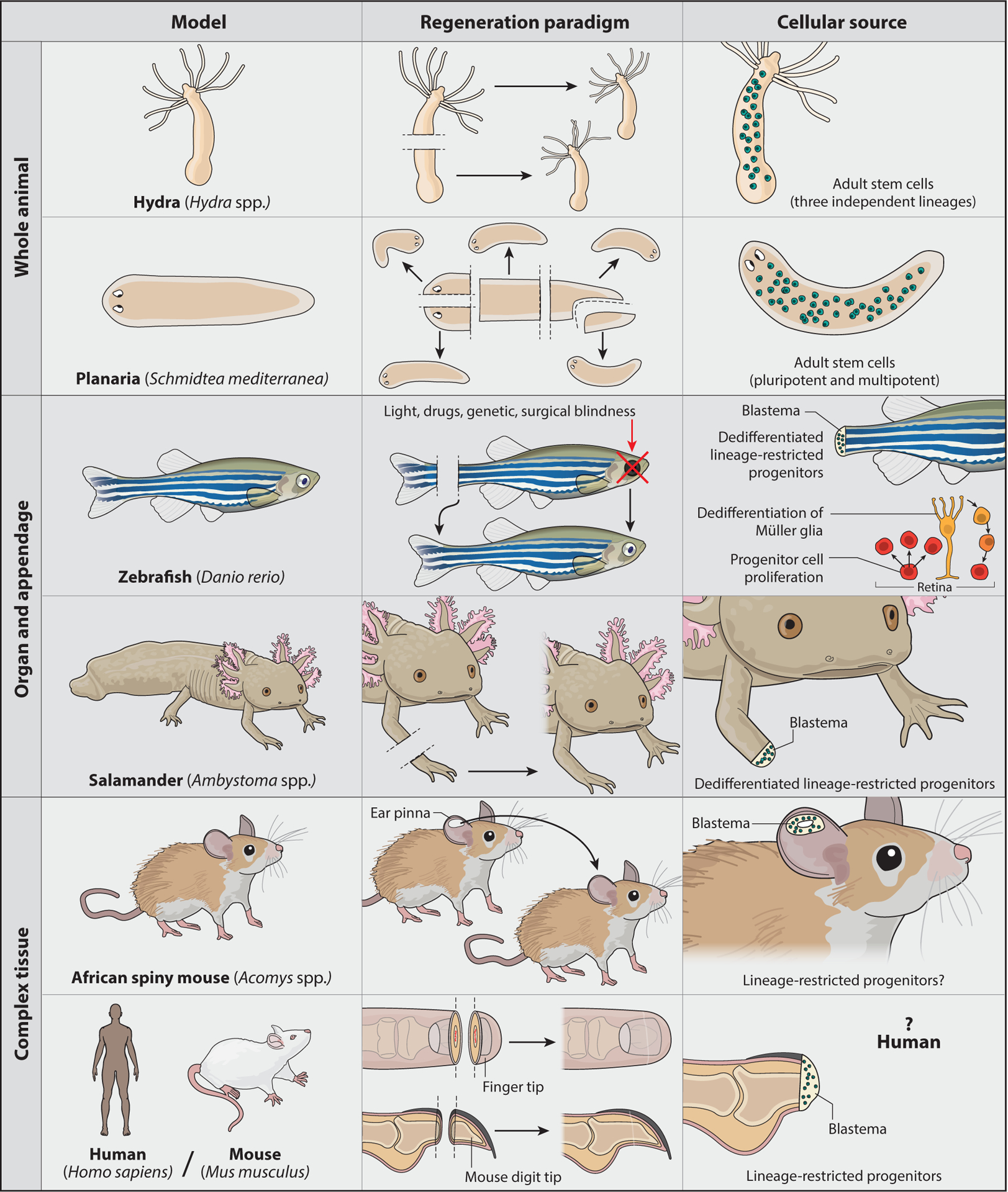
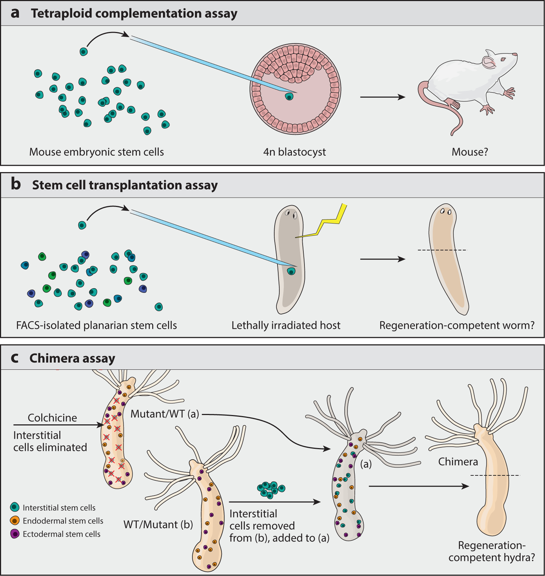
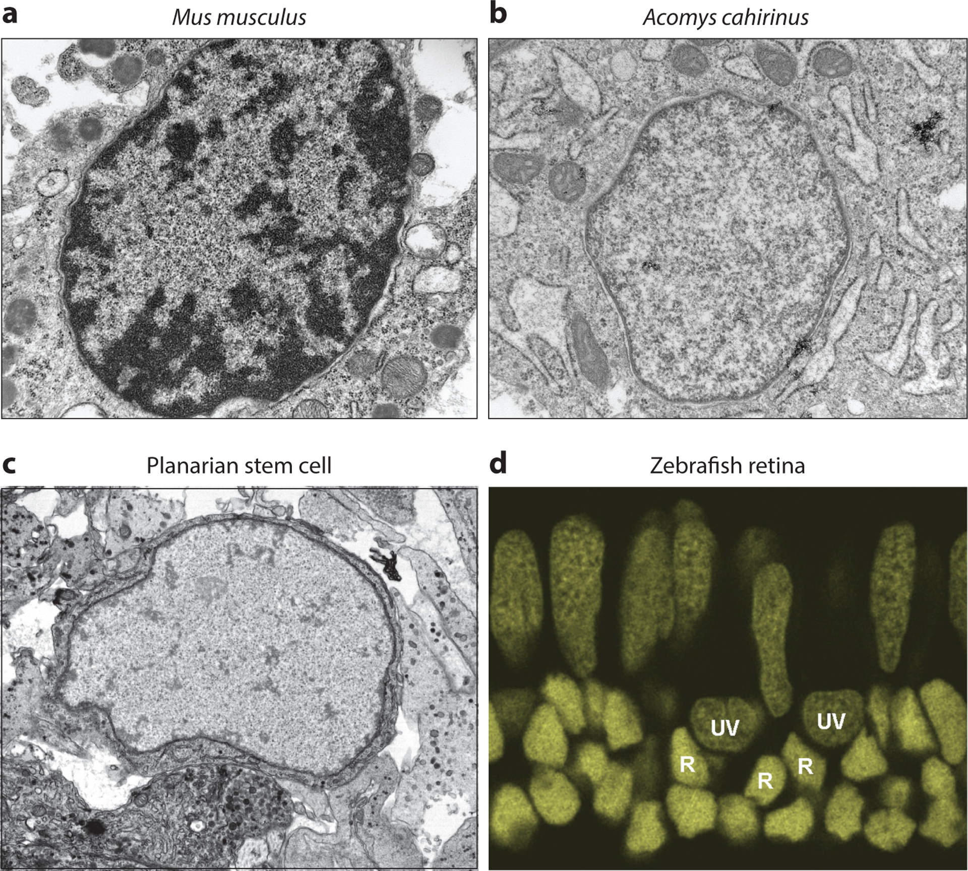
Regeneration is a remarkable phenomenon that has been the subject of awe and bafflement for hundreds of years. Although regeneration competence is found in highly divergent organisms throughout the animal kingdom, recent advances in tools used for molecular and genomic characterization have uncovered common genes, molecular mechanisms, and genomic features in regenerating animals. In this review we focus on what is known about how genome regulation modulates cellular potency during regeneration. We discuss this regulation in the context of complex tissue regeneration in animals, from Hydra to humans, with reference to ex vivo-cultured cell models of pluripotency when appropriate. We emphasize the importance of a detailed molecular understanding of both the mechanisms that regulate genomic output and the functional assays that assess the biological relevance of such molecular characterizations.
Keywords: chromatin; genomic; methylation; planarian; regeneration; stem cell.
Figures
Similar articles
-
On-chip immobilization of planarians for in vivo imaging.Sci Rep. 2014 Sep 17;4:6388. doi: 10.1038/srep06388. Sci Rep. 2014. PMID: 25227263 Free PMC article.
-
Impact of cycling cells and cell cycle regulation on Hydra regeneration.Dev Biol. 2018 Jan 15;433(2):240-253. doi: 10.1016/j.ydbio.2017.11.003. Dev Biol. 2018. PMID: 29291976
-
CBP/p300 homologs CBP2 and CBP3 play distinct roles in planarian stem cell function.Dev Biol. 2021 May;473:130-143. doi: 10.1016/j.ydbio.2021.02.004. Epub 2021 Feb 16. Dev Biol. 2021. PMID: 33607113
-
Why polyps regenerate and we don't: towards a cellular and molecular framework for Hydra regeneration.Dev Biol. 2007 Mar 15;303(2):421-33. doi: 10.1016/j.ydbio.2006.12.012. Epub 2006 Dec 9. Dev Biol. 2007. PMID: 17234176 Review.
-
The cellular basis for animal regeneration.Dev Cell. 2011 Jul 19;21(1):172-85. doi: 10.1016/j.devcel.2011.06.016. Dev Cell. 2011. PMID: 21763617 Free PMC article. Review.
Cited by
-
Molecular regulation of myocardial proliferation and regeneration.Cell Regen. 2021 Apr 6;10(1):13. doi: 10.1186/s13619-021-00075-7. Cell Regen. 2021. PMID: 33821373 Free PMC article. Review.
-
Intestine-enriched apolipoprotein b orthologs are required for stem cell progeny differentiation and regeneration in planarians.Nat Commun. 2022 Jul 1;13(1):3803. doi: 10.1038/s41467-022-31385-2. Nat Commun. 2022. PMID: 35778403 Free PMC article.
-
Cell tip growth underlies injury response of marine macroalgae.PLoS One. 2022 Mar 17;17(3):e0264827. doi: 10.1371/journal.pone.0264827. eCollection 2022. PLoS One. 2022. PMID: 35298494 Free PMC article.
-
An Emerging Frontier in Intercellular Communication: Extracellular Vesicles in Regeneration.Front Cell Dev Biol. 2022 May 11;10:849905. doi: 10.3389/fcell.2022.849905. eCollection 2022. Front Cell Dev Biol. 2022. PMID: 35646926 Free PMC article. Review.
-
A New Protocol of Computer-Assisted Image Analysis Highlights the Presence of Hemocytes in the Regenerating Cephalic Tentacles of Adult Pomacea canaliculata.Int J Mol Sci. 2021 May 9;22(9):5023. doi: 10.3390/ijms22095023. Int J Mol Sci. 2021. PMID: 34065143 Free PMC article.
References
-
- Alibardi L 2010. Morphological and cellular aspects of tail and limb regeneration in lizards. A model system with implications for tissue regeneration in mammals. Adv. Anat. Embryol. Cell Biol 207:1–109 - PubMed
-
- Allis CD, Caparros M-L, Jenuwein T, Reinberg D. 2015. Epigenetics. Cold Spring Harbor, NY: Cold Spring Harbor Laboratory Press. 2nd ed.
Publication types
MeSH terms
Substances
Grants and funding
LinkOut - more resources
Full Text Sources
Medical
