. 2019 Jul 1;142(7):2037-2050.
doi: 10.1093/brain/awz141.

Freezing of gait in Parkinson's disease reflects a sudden derangement of locomotor network dynamics

Affiliations

Freezing of gait in Parkinson's disease reflects a sudden derangement of locomotor network dynamics

Nicoló G Pozzi et al. Brain. .

Abstract

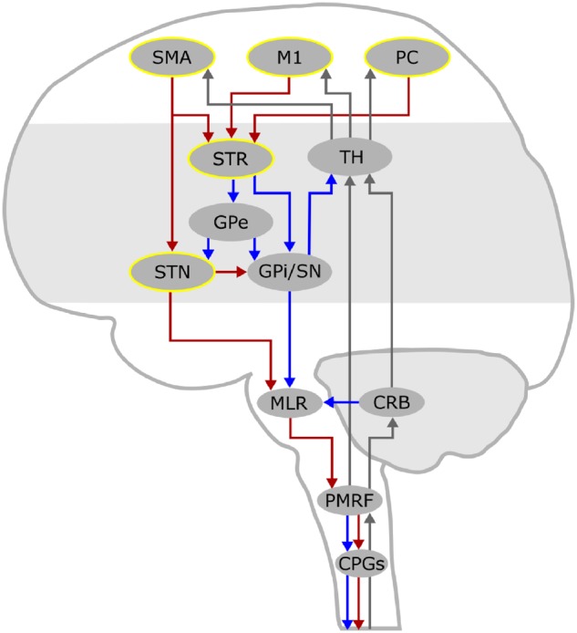
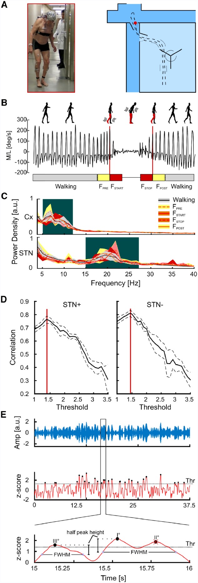
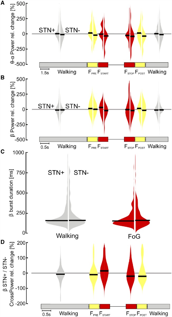
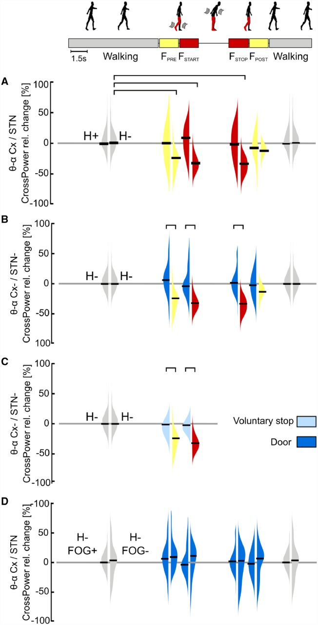
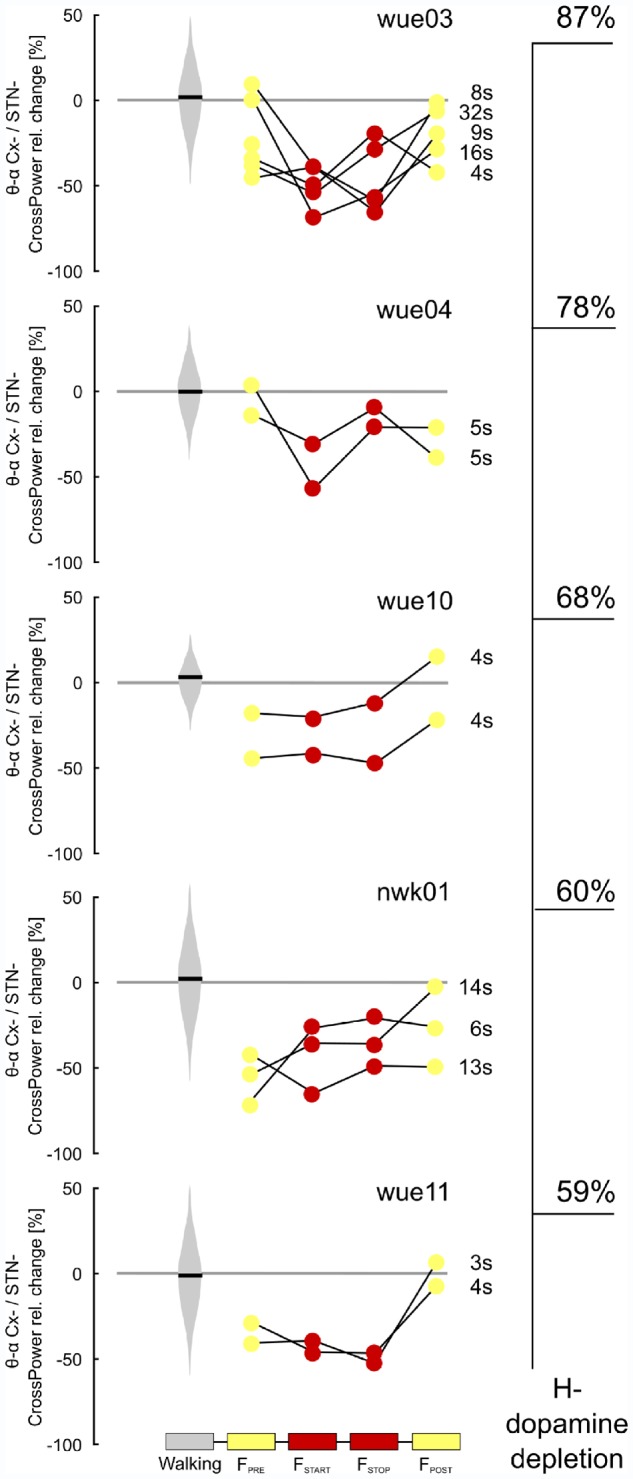
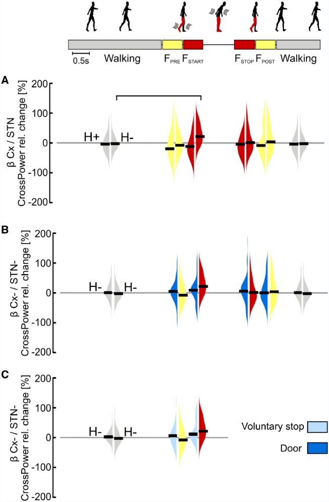
Freezing of gait is a disabling symptom of Parkinson's disease that causes a paroxysmal inability to generate effective stepping. The underlying pathophysiology has recently migrated towards a dysfunctional supraspinal locomotor network, but the actual network derangements during ongoing gait freezing are unknown. We investigated the communication between the cortex and the subthalamic nucleus, two main nodes of the locomotor network, in seven freely-moving subjects with Parkinson's disease with a novel deep brain stimulation device, which allows on-demand recording of subthalamic neural activity from the chronically-implanted electrodes months after the surgical procedure. Multisite neurophysiological recordings during (effective) walking and ongoing gait freezing were combined with kinematic measurements and individual molecular brain imaging studies. Patients walked in a supervised environment closely resembling everyday life challenges. We found that during (effective) walking, the cortex and subthalamic nucleus were synchronized in a low frequency band (4-13 Hz). In contrast, gait freezing was characterized in every patient by low frequency cortical-subthalamic decoupling in the hemisphere with less striatal dopaminergic innervation. Of relevance, this decoupling was already evident at the transition from normal (effective) walking into gait freezing, was maintained during the freezing episode, and resolved with recovery of the effective walking pattern. This is the first evidence for a decoding of the networked processing of locomotion in Parkinson's disease and suggests that freezing of gait is a 'circuitopathy' related to a dysfunctional cortical-subcortical communication. A successful therapeutic approach for gait freezing in Parkinson's disease should aim at directly targeting derangements of neural network dynamics.

Keywords: Parkinson’s disease; basal ganglia; beta oscillations; deep brain stimulation; gait.


Figures

Figure 1
Figure 1
The supraspinal locomotor network. This schematic drawing displays the supraspinal network for locomotor control. Cortical signals convey motor commands to the mesencephalic locomotor regions (MLR) via the basal ganglia, through the striato-pallidal and the striato-subthalamic-pallidal pathways, and via the hyper-direct pathway that directly links the SMA with the STN. Locomotor plans reach the MLR, which represents a cross-point of information coming from the basal ganglia and the cerebellum, and further descend to the pontomedullary reticular formation (PMRF) and to the spinal central pattern generators (CPGs). The investigated nodes are highlighted in yellow. The pathways of feedforward motor commands are displayed as red (activating) and blue (inhibiting) arrows, while those of sensory feedback are displayed as grey arrows. CRB = cerebellum; GPe = globus pallidus pars externa; GPi = globus pallidus pars interna; M1 = primary motor cortex; PC = parietal cortex; SN = substantia nigra; STR = striatum; TH = thalamus.
Figure 2
Figure 2
Experimental set-up, kinematic identification of gait freezing, power spectral densities and β-burst identification analysis. (A) Experimental set-up. The walking path inside and outside the gait laboratory consisted of walking through a turning door (inside the gait laboratory) and two common doors outside, where a representative freezing episode took place (red dot and figure). (B) Kinematic representation of one freezing episode. Representative traces of the ankle angular velocity relative to the medial-lateral axis during (effective) walking and gait freezing. We identified five time frames: (effective) walking is shown as light grey boxes (1.5-s time epochs free of gait freezing), FPRE and FSTOP are the yellow boxes (1.5-s time epochs preceding and following a freezing episode, respectively), and FSTART and FSTOP are shown as red boxes (the first and the last 1.5 s of freezing, respectively). (C) Cortical and STN power spectral densities. The cortical LFPs in the selected regions of interest (SMA, M1 and PC) displayed a bimodal distribution with two distinct activity peaks in the θ- and α-frequency bands. The STN power spectra also showed a bimodal distribution with a small peak at 11 Hz and a prominent peak in the β-frequency band. Shaded areas represent the group level variance computed using the bootstrapping technique (20 repetitions, resampling with replacement) and estimating the confidence intervals between the 5th and 95th percentiles of the bootstrap distributions. The background colour indicates the frequency ranges used for further analyses. (D) β-burst identification. Pearson’s correlation coefficient between average β-amplitude and number of β-peaks above the threshold computed in all 1.5-s walking epochs is reported for the two STN separately (STN+ and STN−). Solid lines are the average correlation curves across subjects. Dashed lines represent the standard error computed with the bootstrap technique. Red lines identify the values used as threshold for β-burst detection. (E) Top: A segment of the wavelet real part (blue line) derived from the wavelet transformed LFPs in the β-peak frequency (20 Hz) of a representative subject is reported. Middle: The wavelet amplitude was z-scored and the β-burst peaks were identified (black dots) and sorted according to their amplitude. We then identified the burst duration with the FWHM method. Bottom: A close view on the identification of burst duration. Starting from the higher peak (peak I°) we found the closest points (blue circles) in which the z-scored wavelet amplitude goes below the peak half amplitude. The time difference between these two points determined the burst duration. Since peak II° was located inside the burst duration of peak I°, we eliminated peak II° and considered these two peaks part of the same burst. STN+ or STN− refers to the side with more and less striatal dopaminergic innervation, respectively.
Figure 3
Figure 3
Subthalamic oscillatory activity and coupling during walking and gait freezing. (A) STN low-frequency power. No difference was found for the relative change of STN low-frequency power during gait freezing with respect to (effective) walking (i.e. zero line) for the two STNs and among them (STN+ versus STN−). (B) STN β-power. The relative change of STN β-power during gait freezing did not differ with respect to (effective) walking (i.e. zero line) for the two STNs and among them (STN+ versus STN−). (C) β-burst duration. No difference was found in the distribution of the β-burst duration during gait freezing with respect to (effective) walking for both STN and among them (STN+ versus STN−). (D) Interhemispheric STN coupling. Differences between the two STNs during gait freezing and (effective) walking did not reach statistical significance. STN+ and STN− refers to the side with more and less striatal dopaminergic innervation, respectively.
Figure 4
Figure 4
Cortical-subthalamic coupling in the low-frequency band. (A) Percentage relative change of cortical-subthalamic low frequency (i.e. θ-α band, 4–13 Hz) coupling during gait freezing versus (effective) walking. During gait freezing (FSTART – FSTOP), the cortex and the STN decoupled selectively in the hemisphere with less striatal dopamine (H−). The decoupling was already evident before gait freezing (FPRE) and vanished with the recovery of a normal locomotor pattern (FPOST). (B) Percentage relative change of cortical-subthalamic low-frequency coupling during gait freezing versus successful passing through a door. The cortex and the STN in H− decoupled only during gait freezing (as in A) and not during successful passing through a door. (C) Percentage relative change of cortical-subthalamic low frequency coupling during gait freezing versus voluntary stop. The cortex and the STN (in H−) decoupled only during gait freezing (as in A) and not during voluntary stop. (D) Percentage relative change of cortical-subthalamic low frequency coupling during passing through a door in subjects with versus without FOG. Subjects with and without FOG showed the same cortical-subthalamic coupling during (effective) walking and successful passing through a door. Cx = cortex (i.e. SMA, M1 and PC); H = hemisphere (H+ and H− refer to the side with more and less striatal dopaminergic innervation, respectively); STN+ and STN− refers to the side with more and less striatal dopaminergic innervation, respectively; FOG+ and FOG− refers to the patients suffering or not suffering from FOG, respectively. The horizontal bars indicate statistical significance (P < 0.05). Of note, the statistical horizontal bars in A are not replicated in B and C, for clarity of the text.
Figure 5
Figure 5
Individual low frequency cortical-subthalamic coupling during gait freezing. Individual percentage relative change of cortical-subthalamic low frequency (i.e. θ-α band, 4–13 Hz) coupling during gait freezing versus (effective) walking for each episode of gait freezing. Subjects are ordered according to the percentage of striatal dopaminergic innervation loss (H−). All subjects and gait freezing episodes showed a similar neurophysiological pattern that was characterized by low frequency cortical-subthalamic decoupling, independent of the duration (in s) of the episode. Cx− = cortex (i.e. SMA, M1 and PC) of the hemisphere with less striatal dopaminergic innervation; H = hemisphere (H− refers to the side with less striatal dopaminergic innervation).
Figure 6
Figure 6
Cortical-subthalamic coupling in the β-frequency. (A) Percentage relative change of cortical-subthalamic β-coupling during gait freezing versus (effective) walking. At gait freezing start (FSTART) the cortical-subthalamic coupling in the β-frequency showed a significant increase in comparison to (effective) walking selectively in the hemisphere with less striatal dopamine (H−). (B) Percentage relative change of cortical-subthalamic β-coupling during gait freezing versus successful passing through a door. The increase in β-coupling between the cortex and the STN (in H−) at gait freezing start (FSTART) was similar to successful passing through a door. (C) Percentage relative change of cortical-subthalamic β-coupling during gait freezing versus voluntary stop. The increase in β-coupling between the cortex and the STN (in H−) did not differ between freezing onset and voluntary stop. Cx = cortex (i.e. SMA, M1 and PC); H = hemisphere (H+ and H− refer to the side with more and less striatal dopaminergic innervation, respectively); STN− refers to the hemisphere with less striatal dopaminergic innervation. Statistical significance is shown with a horizontal bar (P < 0.05). Of note, the statistical horizontal bars in A are not replicated in B and C, for clarity of the text.

References

    1. Anidi CM, O’Day JJ, Anderson RW, Afzal MF, Syrkin-Nikolau J, Velisar A, et al.Neuromodulation targets pathological not physiological beta bursts during gait in Parkinson’s disease. Neurobiol Dis 2018; 120: 107–17. - PMC - PubMed
    1. Arlotti M, Marceglia S, Foffani G, Volkmann J, Lozano AM, Moro E, et al.Eight-hours adaptive deep brain stimulation in patients with Parkinson disease. Neurology 2018. - PMC - PubMed
    1. Arnulfo G, Pozzi NG, Palmisano C, Leporini A, Canessa A, Brumberg J, et al.Phase matters: a role for the subthalamic network during gait. PLoS One 2018; 13: e0198691. - PMC - PubMed
    1. Benda J, Longtin A, Maler L. A synchronization-desynchronization code for natural communication signals. Neuron 2006; 52: 347–58. - PubMed
    1. Brittain JS, Brown P. Oscillations and the basal ganglia: motor control and beyond. Neuroimage 2014; 85: 637–47. - PMC - PubMed

Publication types