

Side Effects of Curcumin: Epigenetic and Antiproliferative Implications for Normal Dermal Fibroblast and Breast Cancer Cells
- PMID: 31505772
- PMCID: PMC6770744
- DOI: 10.3390/antiox8090382
Side Effects of Curcumin: Epigenetic and Antiproliferative Implications for Normal Dermal Fibroblast and Breast Cancer Cells
Abstract
Background: Curcumin is a yellow-orange pigment obtained from the plant Curcuma longa, which is known to exert beneficial effects in several diseases, including cancer. However, at high doses, it may produce toxic and carcinogenic effects in normal cells. In this context, we studied the effects of curcumin on normal human dermal fibroblast (HDF) cells and breast cancer cells (MCF7).
Methods: We used cellular viability and growth assays to evaluate the antiproliferative action of curcumin, analyzed the endogenous glutathione levels, conducted cell cycle, apoptosis, and necrosis analyses, and performed immunodetection of glutathionylated and acetylated H3 histones.
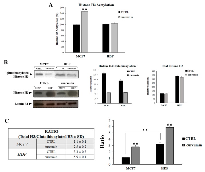
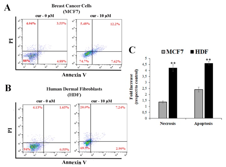
Results: We found that HDFs are more sensitive to curcumin treatment than MCF7 cells, resulting in pronounced arrest of cell cycle progression and higher levels of cellular death. In both cell types, the homeostasis of the redox cellular environment did not change after curcumin treatment; however, significant differences were observed in glutathione (GSH) levels and in S-glutathionylation of H3 histones.
Conclusion: Curcumin administration can potentially confer benefits, but high doses may be toxic. Thus, its use as a dietary supplement or in cancer therapies has a double edge.
Keywords: curcumin; glutathione; histone glutathionylation; human cancer cells; post-translational modifications (PTMs).
Conflict of interest statement
The authors declare no conflict of interest.
Figures


Similar articles
-
The effects of Curcuma longa and curcumin on reproductive systems.Endocr Regul. 2017 Oct 26;51(4):220-228. doi: 10.1515/enr-2017-0024. Endocr Regul. 2017. PMID: 29232190 Review.
-
[Effect of curcumin on acetylation of histone H3 in human lymphoma cell line Raji].Ai Zheng. 2006 May;25(5):582-6. Ai Zheng. 2006. PMID: 16687078 Chinese.
-
Carnosic acid inhibits the growth of ER-negative human breast cancer cells and synergizes with curcumin.Fitoterapia. 2012 Oct;83(7):1160-8. doi: 10.1016/j.fitote.2012.07.006. Epub 2012 Jul 22. Fitoterapia. 2012. PMID: 22828666
-
Curcumin and paclitaxel induce cell death in breast cancer cell lines.Oncol Rep. 2018 Oct;40(4):2381-2388. doi: 10.3892/or.2018.6603. Epub 2018 Jul 26. Oncol Rep. 2018. PMID: 30066930
-
Role of glutathione in the regulation of epigenetic mechanisms in disease.Free Radic Biol Med. 2017 Nov;112:36-48. doi: 10.1016/j.freeradbiomed.2017.07.008. Epub 2017 Jul 10. Free Radic Biol Med. 2017. PMID: 28705657 Review.
Cited by
-
Modulating Effects of Zingiberaceae Phenolic Compounds on Neurotrophic Factors and Their Potential as Neuroprotectants in Brain Disorders and Age-Associated Neurodegenerative Disorders: A Review.Nutrients. 2023 May 30;15(11):2564. doi: 10.3390/nu15112564. Nutrients. 2023. PMID: 37299526 Free PMC article. Review.
-
Curcuminoids as Anticancer Drugs: Pleiotropic Effects, Potential for Metabolic Reprogramming and Prospects for the Future.Pharmaceutics. 2023 May 29;15(6):1612. doi: 10.3390/pharmaceutics15061612. Pharmaceutics. 2023. PMID: 37376060 Free PMC article. Review.
-
Formulation and Evaluation of Microwave-Modified Chitosan-Curcumin Nanoparticles-A Promising Nanomaterials Platform for Skin Tissue Regeneration Applications Following Burn Wounds.Polymers (Basel). 2020 Nov 6;12(11):2608. doi: 10.3390/polym12112608. Polymers (Basel). 2020. PMID: 33171959 Free PMC article.
-
Protection of Polyphenols against Glyco-Oxidative Stress: Involvement of Glyoxalase Pathway.Antioxidants (Basel). 2020 Oct 16;9(10):1006. doi: 10.3390/antiox9101006. Antioxidants (Basel). 2020. PMID: 33081239 Free PMC article.
-
A Combination of Curcumin and Lactobacillus rhamnosus GG Inhibits Viability and Induces Apoptosis in SCC-9 Human Oral Squamous Cell Carcinoma Cells.J Evid Based Integr Med. 2024 Jan-Dec;29:2515690X241258369. doi: 10.1177/2515690X241258369. J Evid Based Integr Med. 2024. PMID: 38778767 Free PMC article.
References
-
- Bhattacharyya S., Mandal D., Sen G.S., Pal S., Banerjee S., Lahiry L., Finke J.H., Tannenbaum C.S., Das T., Sa G. Tumor-induced oxidative stress perturbs nuclear factor-kappaB activity-augmenting tumor necrosis factor-alpha-mediated T-cell death: protection by curcumin. Cancer Res. 2007;67:362–370. doi: 10.1158/0008-5472.CAN-06-2583. - DOI - PubMed
LinkOut - more resources
Full Text Sources
Miscellaneous
