Review
. 2019 Sep 9;20(18):4431.
doi: 10.3390/ijms20184431.

Involvement of Novel Adipokines, Chemerin, Visfatin, Resistin and Apelin in Reproductive Functions in Normal and Pathological Conditions in Humans and Animal Models

Affiliations
Review

Involvement of Novel Adipokines, Chemerin, Visfatin, Resistin and Apelin in Reproductive Functions in Normal and Pathological Conditions in Humans and Animal Models

Anthony Estienne et al. Int J Mol Sci. .

Abstract

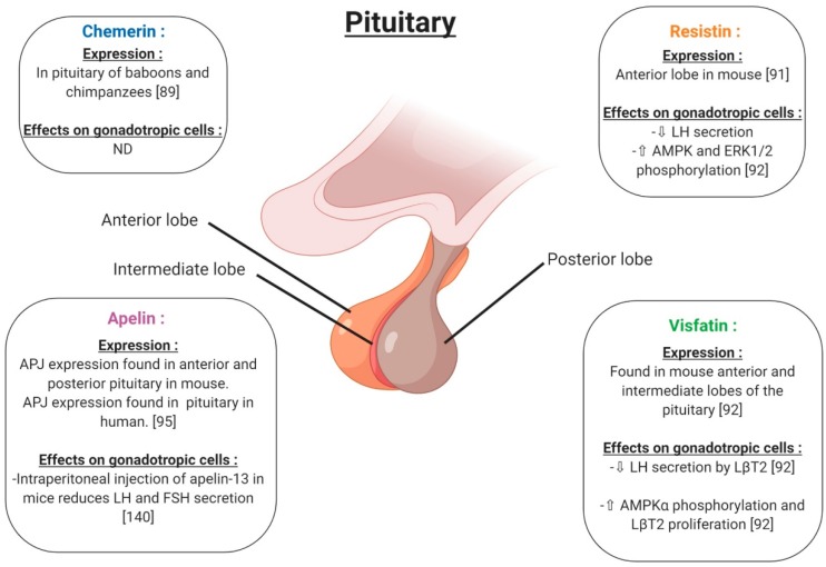
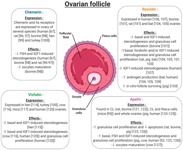
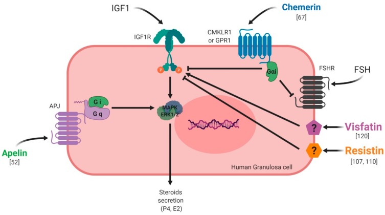
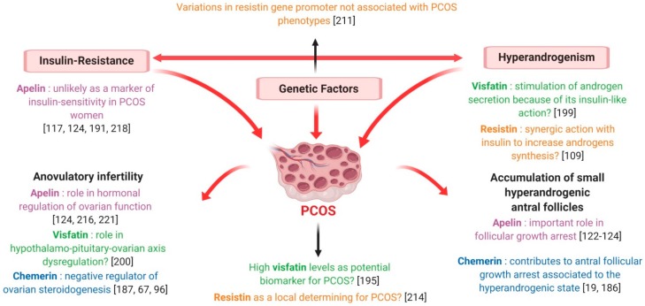
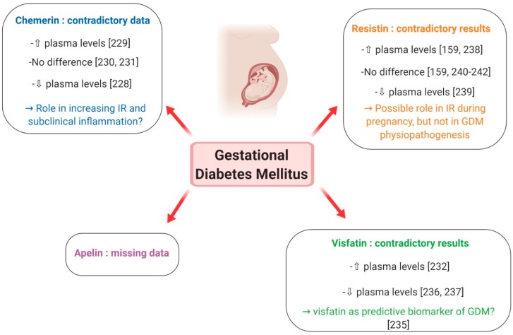
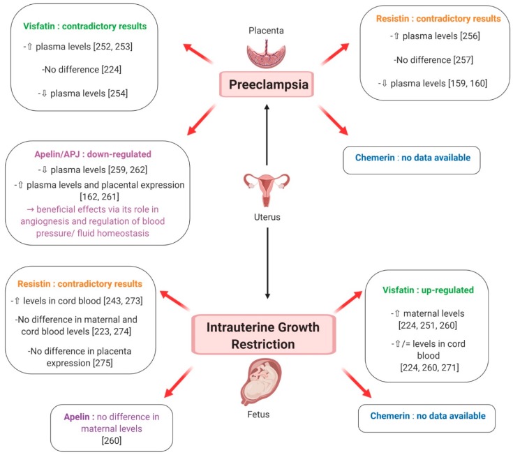
It is well known that adipokines are endocrine factors that are mainly secreted by white adipose tissue. Their central role in energy metabolism is currently accepted. More recently, their involvement in fertility regulation and the development of some reproductive disorders has been suggested. Data concerning the role of leptin and adiponectin, the two most studied adipokines, in the control of the reproductive axis are consistent. In recent years, interest has grown about some novel adipokines, chemerin, visfatin, resistin and apelin, which have been found to be strongly associated with obesity and insulin-resistance. Here, we will review their expression and role in male and female reproduction in humans and animal models. According to accumulating evidence, they could regulate the secretion of GnRH (Gonadotropin-Releasing Hormone), gonadotropins and steroids. Furthermore, their expression and that of their receptors (if known), has been demonstrated in the human and animal hypothalamo-pituitary-gonadal axis. Like leptin and adiponectin, these novel adipokines could thus represent metabolic sensors that are able to regulate reproductive functions according to energy balance changes. Therefore, after investigating their role in normal fertility, we will also discuss their possible involvement in some reproductive troubles known to be associated with features of metabolic syndrome, such as polycystic ovary syndrome, gestational diabetes mellitus, preeclampsia and intra-uterine growth retardation in women, and sperm abnormalities and testicular pathologies in men.

Keywords: adipose tissue; gestational diabetes; ovary; polycystic ovary syndrome; preeclempsia; testicular pathologies; testis.


Conflict of interest statement

The authors declare no conflict of interest. The funders had no role in the design of the study; in the collection, analyses, or interpretation of data; in the writing of the manuscript, or in the decision to publish the results.

Figures

Figure 1
Figure 1
Structure and specific forms of human chemerin. The initial preprochemerin and its different products are processed by different proteases related to inflammation. The signal peptide (purple) is cleaved prior to secretion. Then, the C-terminus is cleaved by different proteases giving several active isoforms such as chemerin F156, chemerin S157 and chemerin K158.
Figure 2
Figure 2
Structure and specific forms of human visfatin. Human visfatin can be found under the intra-cellular form of nicotinamide phosphoribosyltransferase (iNAMPT) having an enzymatic role to produce NAD+ (nicotinamide adenine dinucleotide), and under the extracellular form (eNAMPT) with the same role. Visfatin acts also as a cytokine that could act on target cells. NAMPT catalyzes the reaction between nicotinamide and 5-phosphoribosyl-1-pyrophosphate (PRPP) to yield nicotinamide mononucleotide (NMN), an intermediate in the biosynthesis of NAD+.
Figure 3
Figure 3
Structure and specific forms of human resistin. Human resistin is composed by a signal peptide (purple), a variable region (green) and a C-terminal domain. Resistin can be found under the monomeric form and can form dimeric and trimeric proteins thanks to disulfide bridges. Then, disulfide and non-disulfide bridges can be involved in the formation of the hexameric protein.
Figure 4
Figure 4
Structure and specific forms of human apelin. Human apelin is first of all found under the preproapelin form (77 amino acids) that will be cleaved by endopeptidases acting on basic amino-acid-rich regions giving the proapelin (55 amino-acids) and then the other tissue-dependent active isoforms (36, 17, 13 and Pyr-13 amino-acids). PCSK3 (proprotein convertase subtilisin/kexin 3) is involved in the cleavage of proapelin to apelin-13.
Figure 5
Figure 5
Receptors used by chemerin, visfatin, resistin and apelin for their signaling pathways. chemerin can bind three different receptors that are CMKLR1, GPR1 and CCRL2. CMKLR1 and GPR1 are coupled with intracellular Gαi proteins. The last receptor doesn’t have any active signaling pathway identified until now suggesting a putative role of co-receptor for CCRL2. Apelin has its own receptor called APJ coupled with intracellular Gq and Gαi proteins. Resistin could bind receptors such as CAP-1, ROR-1 and TLR-4 but it has to be confirmed. No receptor for visfatin has been identified until now.
Figure 6
Figure 6
Expression and effects on GnRH (Gonadotropin-Releasing Hormone) release of chemerin, visfatin, resistin and apelin in hypothalamus. ⇧ Increase/stimulation. ⇩ Decrease/inhibition. ND: not determined. CSF: cerebrospinal fluid.
Figure 7
Figure 7
Expression and effects on gonadotropic cells of chemerin, visfatin, resistin and apelin in pituitary. ⇧ Increase/stimulation. ⇩ Decrease/inhibition. ND: not determined.
Figure 8
Figure 8
Expression and effects of chemerin, visfatin, resistin and apelin on the ovarian follicle. ⇧ Increase/stimulation. ⇩ Decrease/inhibition.
Figure 9
Figure 9
Effects of chemerin, visfatin, resistin and apelin on steroidogenesis in vitro in primary human granulosa cells. Chemerin and resistin are known to inhibit IGF-1 (Insulin Like Growth Factor 1)-induced steroidogenesis whereas visfatin and apelin exert stimulatory effects.
Figure 10
Figure 10
Expression and effects of chemerin, visfatin, resistin and apelin on testicular function. ⇧ Increase/stimulation. ⇩ Decrease/inhibition.
Figure 11
Figure 11
Effects of chemerin, visfatin, resistin and apelin on ovarian physiology, plasma and adipose tissue in polycystic ovarian syndrome (PCOS) as compared to control patients. ⇧ Increase/stimulation. ⇩ Decrease/inhibition.
Figure 12
Figure 12
Description of PCOS syndrome and possible involvement of chemerin, visfatin, resistin and apelin in this syndrome.
Figure 13
Figure 13
Putative involvement of plasma levels of chemerin, visfatin, resistin and apelin in gestational diabetes mellitus. ⇧ Increase/stimulation. ⇩ Decrease/inhibition.
Figure 14
Figure 14
Putative involvement of plasma levels of chemerin, visfatin, resistin and apelin in preeclampsia and intrauterine growth restriction. ⇧ Increase/stimulation. ⇩ Decrease/inhibition.

Similar articles

Cited by

References

    1. Wade G.N., Schneider J.E. Metabolic fuels and reproduction in female mammals. Neurosci. Biobehav. Rev. 1992;16:235–272. doi: 10.1016/S0149-7634(05)80183-6. - DOI - PubMed
    1. Schneider J.E. Energy balance and reproduction. Physiol. Behav. 2004;81:289–317. doi: 10.1016/j.physbeh.2004.02.007. - DOI - PubMed
    1. Scheja L., Heeren J. The endocrine function of adipose tissues in health and cardiometabolic disease. Nat. Rev. Endocrinol. 2019;15:507–524. doi: 10.1038/s41574-019-0230-6. - DOI - PubMed
    1. Ntaios G., Gatselis N.K., Makaritsis K., Dalekos G.N. Adipokines as mediators of endothelial function and atherosclerosis. Atherosclerosis. 2013;227:216–221. doi: 10.1016/j.atherosclerosis.2012.12.029. - DOI - PubMed
    1. Trujillo M.E., Scherer P.E. Adipose tissue-derived factors: Impact on health and disease. Endocr. Rev. 2006;27:762–778. doi: 10.1210/er.2006-0033. - DOI - PubMed