Review
. 2019 Sep 9;7(9):336.
doi: 10.3390/microorganisms7090336.

The Intestine of Drosophila melanogaster: An Emerging Versatile Model System to Study Intestinal Epithelial Homeostasis and Host-Microbial Interactions in Humans

Affiliations
Review

The Intestine of Drosophila melanogaster: An Emerging Versatile Model System to Study Intestinal Epithelial Homeostasis and Host-Microbial Interactions in Humans

Florence Capo et al. Microorganisms. .

Abstract

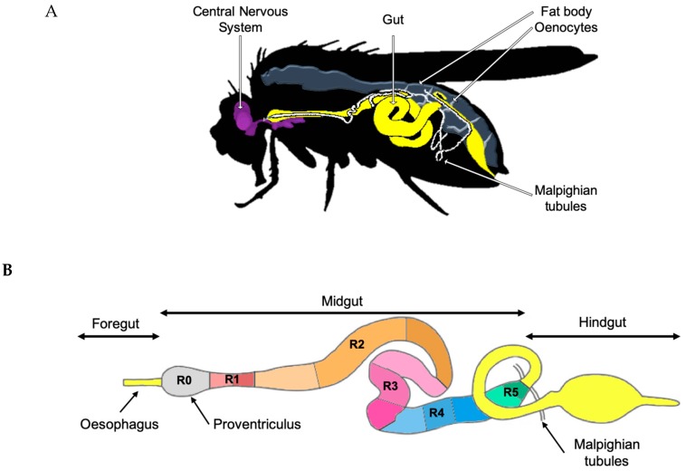
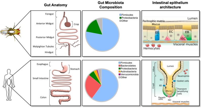
In all metazoans, the intestinal tract is an essential organ to integrate nutritional signaling, hormonal cues and immunometabolic networks. The dysregulation of intestinal epithelium functions can impact organism physiology and, in humans, leads to devastating and complex diseases, such as inflammatory bowel diseases, intestinal cancers, and obesity. Two decades ago, the discovery of an immune response in the intestine of the genetic model system, Drosophila melanogaster, sparked interest in using this model organism to dissect the mechanisms that govern gut (patho) physiology in humans. In 2007, the finding of the intestinal stem cell lineage, followed by the development of tools available for its manipulation in vivo, helped to elucidate the structural organization and functions of the fly intestine and its similarity with mammalian gastrointestinal systems. To date, studies of the Drosophila gut have already helped to shed light on a broad range of biological questions regarding stem cells and their niches, interorgan communication, immunity and immunometabolism, making the Drosophila a promising model organism for human enteric studies. This review summarizes our current knowledge of the structure and functions of the Drosophila melanogaster intestine, asserting its validity as an emerging model system to study gut physiology, regeneration, immune defenses and host-microbiota interactions.

Keywords: Drosophila melanogaster; host-pathogen/commensal interactions; immunometabolism; inflammatory bowel disease; innate immunity; intestinal epithelium; microbiota; midgut; small intestine.


Conflict of interest statement

The authors declare no conflict of interests.

Figures

Figure 1
Figure 1
Adult Drosophila melanogaster anatomy. (A) The Drosophila melanogaster model system contains tissues that functionally correspond to most essential human organs: central nervous system, gastrointestinal system, adipose tissue and the liver (synergic function of fat body and oenocytes) and kidneys (Malpighian tubules). (B) The Drosophila melanogaster adult foregut, midgut, and hindgut. The midgut consists of six major anatomical regions (R0–R5) which are further subdivided into 14 color-coded sub-regions (for example, R2 is subdivided into three orange sub-regions) according to morphometric, histochemical and transcriptomic data.
Figure 2
Figure 2
Comparisons between Drosophila melanogaster and human gut anatomy, gut bacterial microbiota, and intestinal epithelium. Left panel: Gut anatomy of Drosophila melanogaster and humans. Drosophila midgut and hindgut are functional analogs of the human small intestine and colon, respectively (extracted and modified from Figure 1 of [20]). Middle panel: Taxonomical distribution data for top phyla in Drosophila melanogaster and humans (extracted from Figure 1 of [20]). Right panel: Intestinal epithelium is surrounded by visceral muscles and composed of intestinal stem cells (ISCs) in red, undifferentiated ISC daughters in green (enteroblasts (EBs)), enterocytes (ECs) in beige and enteroendocrine cells (EEs) in blue. Under normal conditions, the gut microbiota is localized within the lumen. Barriers such as the peritrophic membrane within Drosophila melanogaster and the mucus within humans prevent direct contact between intestinal epithelial cells and gut bacteria.
Figure 3
Figure 3
Intracellular peptidoglycan (PGN) is detected by intracellular receptors and activates the NF-κB signaling pathway in Drosophila melanogaster and human enterocytes. Intracellular PGN is detected by the PGRP-LE receptor in Drosophila melanogaster (A) or by NOD receptors in human (B). PGN-receptor interaction triggers the NF-κB signaling cascade (Imd pathway in Drosophila), which results in the production of antimicrobial peptides (AMPs). The orthologous proteins are represented by the same shape, color and position in the pathways.
Figure 4
Figure 4
The dual oxidase (DUOX) pathway and reactive oxygen species (ROS) regulation. (A) Diagram of the conserved DUOX protein domains in Drosophila melanogaster and humans. In Drosophila, the peroxidase homology domain of DUOX converts H2O2 into HOCl in the presence of chloride. DUOX-dependent H2O2 molecules are eliminated by immune-regulated catalase (IRC) activity. In humans, DUOX-dependent H2O2 is used for the oxidative conversion of SCN to OSCN by the enzymatic action of lactoperoxidase in the mucosal fluids (extracted and modified from Figure 1A of [139]). (B) In Drosophila enterocytes, peroxisomes integrate and modulate the stress and immune pathways to maintain enteric homeostasis, mount host defense in the gut, and promote gut renewal. These processes are impaired in the presence of dysfunctional peroxisomes and lead to an intracellular accumulation of ROS and free fatty acids (FAAs), and cell death [149].

Similar articles

Cited by

References

    1. Raz E. Mucosal immunity: Aliment and ailments. Mucosal Immunol. 2009;3:4–7. doi: 10.1038/mi.2009.123. - DOI - PubMed
    1. Kiela P.R., Ghishan F.K. Physiology of Intestinal Absorption and Secretion. Best Pract. Res. Clin. Gastroenterol. 2016;30:145–159. doi: 10.1016/j.bpg.2016.02.007. - DOI - PMC - PubMed
    1. Satsu H. Molecular and cellular studies on the absorption, function, and safety of food components in intestinal epithelial cells. Biosci. Biotechnol. Biochem. 2017;81:419–425. doi: 10.1080/09168451.2016.1259552. - DOI - PubMed
    1. Okumura R., Takeda K. Roles of intestinal epithelial cells in the maintenance of gut homeostasis. Exp. Mol. Med. 2017;49:e338. doi: 10.1038/emm.2017.20. - DOI - PMC - PubMed
    1. Woods S.C., Seeley R.J., Porte D., Schwartz M.W. Signals that regulate food intake and energy homeostasis. Science. 1998;280:1378–1383. doi: 10.1126/science.280.5368.1378. - DOI - PubMed

LinkOut - more resources