. 2019 Sep 27;16(158):20190376.
doi: 10.1098/rsif.2019.0376. Epub 2019 Sep 11.

Robust synchronization of the cell cycle and the circadian clock through bidirectional coupling

Affiliations

Robust synchronization of the cell cycle and the circadian clock through bidirectional coupling

Jie Yan et al. J R Soc Interface. .

Abstract

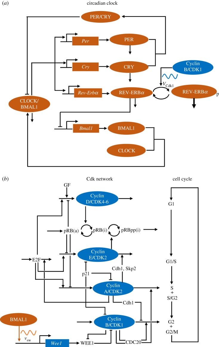
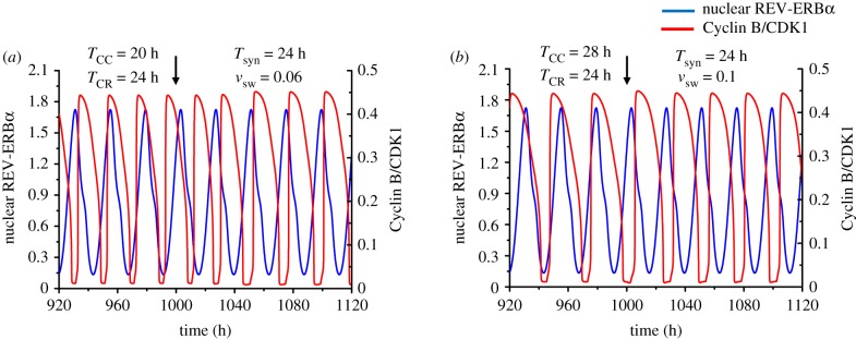
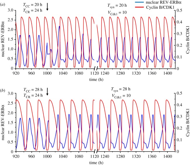
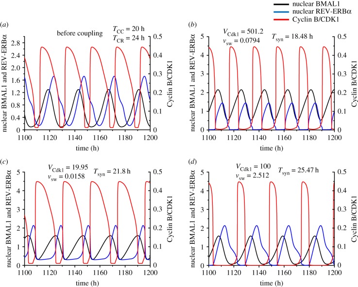
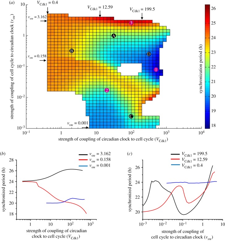
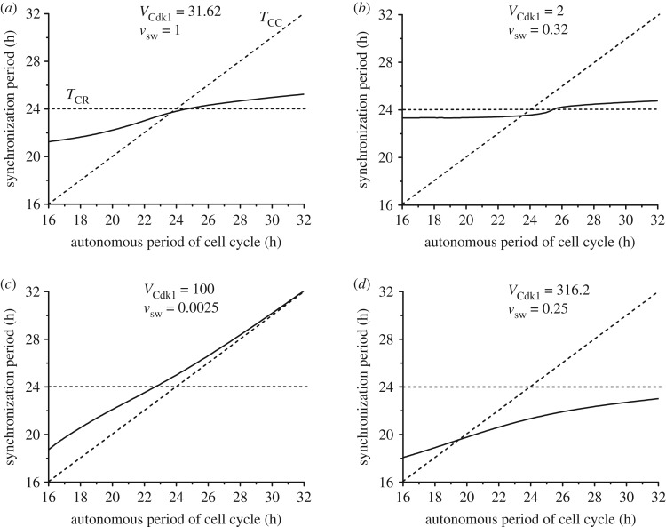
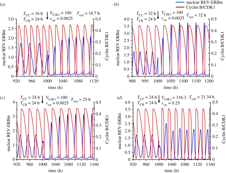
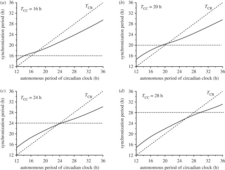
The cell cycle and the circadian clock represent major cellular rhythms, which appear to be coupled. Thus the circadian factor BMAL1 controls the level of cell cycle proteins such as Cyclin E and WEE1, the latter of which inhibits the kinase CDK1 that governs the G2/M transition. In reverse the cell cycle impinges on the circadian clock through direct control by CDK1 of REV-ERBα, which negatively regulates BMAL1. These observations provide evidence for bidirectional coupling of the cell cycle and the circadian clock. By merging detailed models for the two networks in mammalian cells, we previously showed that unidirectional coupling to the circadian clock can entrain the cell cycle to 24 or 48 h, depending on the cell cycle autonomous period, while complex oscillations occur when entrainment fails. Here we show that the reverse unidirectional coupling via phosphorylation of REV-ERBα or via mitotic inhibition of transcription, both controlled by CDK1, can elicit entrainment of the circadian clock by the cell cycle. We then determine the effect of bidirectional coupling of the cell cycle and circadian clock as a function of their relative coupling strengths. In contrast to unidirectional coupling, bidirectional coupling markedly reduces the likelihood of complex oscillations. While the two rhythms oscillate independently as long as both couplings are weak, one rhythm entrains the other if one of the couplings dominates. If the couplings in both directions become stronger and of comparable magnitude, the two rhythms synchronize, generally at an intermediate period within the range defined by the two autonomous periods prior to coupling. More surprisingly, synchronization may also occur at a period slightly below or above this range, while in some conditions the synchronization period can even be much longer. Two or even three modes of synchronization may sometimes coexist, yielding examples of birhythmicity or trirhythmicity. Because synchronization readily occurs in the form of simple periodic oscillations over a wide range of coupling strengths and in the presence of multiple connections between the two oscillatory networks, the results indicate that bidirectional coupling favours the robust synchronization of the cell cycle and the circadian clock.

Keywords: cell cycle; circadian clock; coupled oscillators; synchronization.


Conflict of interest statement

We declare we have no competing interests.

Figures

Figure 1.
Figure 1.
Schematic of the models for (a) the circadian clock and (b) the cell cycle in mammalian cells. In (a), the circadian clock network (in orange) involves the negative autoregulation of the Per and Cry genes, via the inhibition of the activators BMAL1 and CLOCK by the PER and CRY proteins. An additional negative feedback on Bmal1 expression is mediated by the REV-ERBα protein, which is itself induced by CLOCK/BMAL1. These feedback regulations are responsible for the onset of circadian oscillations in the network (see [–22] for reviews, and [24] for further details on the model). The cell cycle controls the circadian clock through several interactions, only one of which is shown: the phosphorylation by CDK1 (of maximum rate VCdk1) of REV-ERBα, which enhances the degradation of this protein. In (b), the CDK network (in blue) that governs the dynamics of the mammalian cell cycle consists of four CDK modules, centred on the complexes Cyclin D/CDK4–6, Cyclin E/CDK2, Cyclin A/CDK2 and Cyclin B/CDK1, which control, respectively, the progression along the G1, S and G2 phases and the G2/M transition [23], as shown on the right part of (b). Also shown in this scheme are some of the important protein factors involved in regulation of the CDK network: growth factors (GF), the retinoblastoma protein, non-phosphorylated (pRB) or inactivated through one (pRBp) or multiple phosphorylations (pRBpp) by CDK1 and CDK2 (a, active; i, inactive); the transcription factor E2F; the CDK inhibitor p21; the proteins Cdh1, Skp2 and CDC20 involved in cyclin degradation; and the kinase WEE1, which inhibits CDK1. Only some of the main regulatory interactions are shown in this simplified scheme. The regulatory design of the CDK network is such that each CDK module activates the next module and inhibits the previous one. Such a regulation results in the repetitive, ordered, transient activation of the four CDK modules that control the successive phases of the cell cycle. The circadian clock controls the cell cycle through several interactions, only one of which is shown: the induction by BMAL1 of the expression (at a rate vsw) of the gene Wee1, which codes for the inhibitory kinase WEE1. The models for the cell cycle and circadian clock contain 41 and 19 variables, respectively. The models for the circadian clock and the cell cycle are described in more detail in [24] and [25], respectively. (Online version in colour.)
Figure 2.
Figure 2.
Unidirectional coupling of the cell cycle to the circadian clock. The time series show the entrainment of the cell cycle by the circadian clock when the autonomous period of the cell cycle (TCC) is 20 h (a) or 28 h (b), while the autonomous period of the circadian rhythm (TCR) is 24 h. In both cases the cell cycle can be entrained to oscillate with a period of 24 h when it is unidirectionally coupled to the circadian clock via induction of Wee1 by BMAL1, starting at the time marked by the vertical arrow. Before coupling begins, the time series show the cell cycle and the circadian clock oscillating independently at their autonomous period. The strength of coupling of the cell cycle to the circadian clock, vsw, is equal to 0.06 µMh−1 in (a) and 0.1 µMh−1 in (b). The scaling parameter eps used to fix the cell cycle period is equal to 21.58 in (a) and 15.35 in (b). The blue curve represents the time evolution of nuclear REV-ERBα (a circadian variable) while the red curve represents the time evolution of Cyclin B/CDK1 (a cell cycle variable). For this and subsequent figures, parameter values are given in the electronic supplementary material. (Online version in colour.)
Figure 3.
Figure 3.
Unidirectional coupling of the circadian clock to the cell cycle. The time series show the entrainment of the circadian clock by the cell cycle when the autonomous period of the latter (TCC) is 20 h (a) or 28 h (b), while the autonomous period of the circadian rhythm (TCR) is 24 h. In both cases the circadian clock is entrained to oscillate at the cell cycle period upon unidirectional coupling via phosphorylation of REV-ERBα by Cyclin B/CDK1, starting at the time marked by the vertical arrow (t = 1000 h). Before coupling begins, the time series show the cell cycle and the circadian clock oscillating independently at their autonomous period. The strength of coupling of the circadian clock to the cell cycle, VCdk1, is equal to 10 nMh−1 in (a) and (b), while the scaling parameter eps is 21.58 in (a) and 15.35 in (b). The blue curve represents the time evolution of nuclear REV-ERBα while the red curve represents the time evolution of Cyclin B/CDK1. In both panels time is interrupted between 1120 h and 1220 h to reduce the number of transients shown after the onset of coupling. (Online version in colour.)
Figure 4.
Figure 4.
Bidirectional coupling of the circadian clock and the cell cycle, through BMAL1 induction of Wee1 and REV-ERBα phosphorylation by CDK1. Before coupling, the cell cycle and the circadian clock oscillate independently with an autonomous period of 20 h and 24 h, respectively (a). Upon bidirectional coupling at t = 1110 h (vertical arrows), depending on the strengths of coupling, the cell cycle and the circadian clock can synchronize at a period shorter than 20 h (b), between 20 and 24 h (c), or longer than 24 h (d). In (b) where the synchronization period is 18.48 h, the coupling strength of the cell cycle to the circadian clock (vsw, in μMh−1) is 0.0794 and the reverse coupling strength (VCdk1, in nMh−1) is 501.2. In (c) where the synchronization period is 21.8 h, vsw = 0.0158 and VCdk1 = 19.95. In (d) the synchronization period is 25.47 h, vsw = 2.512 and VCdk1 = 100. Curves in black, blue and red represent the time evolution of nuclear BMAL1, nuclear REV-ERBα and Cyclin B/CDK1, respectively. The transients in (b)–(d) last for one or two cycles. The data in (b), (c) and (d) correspond, respectively, to the points marked 1, 2 and 3 in figure 5a. (Online version in colour.)
Figure 5.
Figure 5.
Bidirectional coupling: dependence of the synchronization period on the relative strengths of coupling of the cell cycle and the circadian clock. The cell cycle is coupled to the circadian clock through BMAL1 induction of Wee1 while the circadian clock is linked to the cell cycle through REV-ERBα phosphorylation by CDK1. (a) Heat map showing how the period of synchronization varies as a function of the strength of coupling of the cell cycle to the circadian clock (vsw, in μMh−1) and of the strength of coupling of the circadian clock to the cell cycle (VCdk1, in nMh−1). The diagram is established for an autonomous period TCC = 20 h for the cell cycle and TCR = 24 h for the circadian clock. Coloured regions indicate synchronization in the form of simple periodic oscillations; the colour code for the synchronization period, on the right, ranges from 17.8 to 26.3 h. Points marked 1, 2 and 3 refer to the synchronized oscillations shown in figure 4b, c and d, respectively. Points marked A, B, C and D correspond, respectively, to the situations considered in figure 6ad. (b) Three horizontal sections through the heat map in (a) as a function of the strength of coupling of the circadian clock to the cell cycle, VCdk1, at decreasing values of the strength of coupling of the cell cycle to the circadian clock, vsw. For vsw = 3.162, or 0.158, as VCdk1 increases, the period of synchronization ranges, respectively, from 24.1 h to 26.3 h (black curve) or from 24 h to 18.1 h (red curve), while it remains close to 20 h when vsw = 0.001 (blue curve). (c) Three vertical sections through the heat map in (a) as a function of the strength of coupling of the cell cycle to the circadian clock, vsw, at decreasing values of the strength of coupling of the circadian clock to the cell cycle, VCdk1. For VCdk1 = 199.5 or 12.59, the synchronization period ranges from 19.65 to 26.23 h (black curve) or from 20 to 25.39 h (red curve) as vsw increases, while it remains close to 24 h when VCdk1 = 0.4 (blue curve). (Online version in colour.)
Figure 6.
Figure 6.
Bidirectional coupling: dependence of the synchronization period on the autonomous period of the cell cycle, TCC. As in figures 4 and 5, the cell cycle is coupled to the circadian clock through BMAL1 induction of Wee1 while the circadian clock is linked to the cell cycle through REV-ERBα phosphorylation by CDK1. The autonomous period of the circadian clock TCR is fixed at 24 h, while the autonomous period of the cell cycle increases from 16 to 32 h (by changing the scaling parameter eps from 26.9 to 13.4; see Section 1 in the electronic supplementary material). The values of the coupling strengths VCdk1 (in nMh−1) and vsw (in μMh−1) are of comparable magnitude in (a) (VCdk1 = 31.62, vsw = 1), while in (b) the coupling of the circadian clock to the cell cycle is weaker (VCdk1 = 2, vsw = 0.32). In (c) the coupling of the circadian clock to the cell cycle is much stronger than the reverse coupling (VCdk1 = 100, vsw = 0.0025), as in (d) where the curve was obtained for larger values of the two coupling strengths (VCdk1 = 316.2, vsw = 0.25). In each panel the synchronization period Tsyn (solid line) increases gradually with TCC. The horizontal and diagonal dashed lines correspond, respectively, to Tsyn = TCR and Tsyn = TCC. All synchronized oscillations are of the simple periodic type with one peak of each variable per period. The data in (a), (b), (c), and (d) correspond, respectively, to the points marked A, B, C and D in figure 5a.
Figure 7.
Figure 7.
Bidirectional coupling of the circadian clock and the cell cycle for different values of the cell cycle autonomous period, TCC. As in figures 4–6, the cell cycle is coupled to the circadian clock through BMAL1 induction of Wee1 while the circadian clock is linked to the cell cycle through REV-ERBα phosphorylation by CDK1. (a) Before coupling, the cell cycle and the circadian clock oscillate independently with an autonomous period of 16 h and 24 h, respectively. Upon coupling, with VCdk1 = 100 (in nMh−1) and vsw = 0.0025 (in μMh−1), the two oscillators synchronize at a period of 18.7 h. (b) When the autonomous period of the cell cycle is increased to 32 h, the cell cycle and the circadian clock synchronize after coupling at a period of 32 h. (cd) When the autonomous period is 24 h for both the cell cycle and the circadian clock, the synchronized period upon coupling can be longer or shorter than 24 h, e.g. 25 h in (c) or 21.34 h in (d). The coupling strengths are VCdk1 = 100, vsw = 0.0025 in (c), and VCdk1 = 316.2 and vsw = 0.25 in (d). The scaling parameter eps is equal to 26.9 in (a), 13.4 in (b), and 17.9 in (c) and (d). (Online version in colour.)
Figure 8.
Figure 8.
Bidirectional coupling: dependence of the synchronization period on the autonomous period of the circadian clock, TCR. As in figures 4–7, the cell cycle is coupled to the circadian clock via BMAL1 induction of Wee1 while the circadian clock is linked to the cell cycle through REV-ERBα phosphorylation by CDK1 (see Sections 2 and 3 in the electronic supplementary material). The coupling strengths are fixed at the values VCdk1 = 31.62 and vsw = 1 considered in figure 6a. The autonomous period of the circadian clock increases from 12 to 36 h by progressively changing the scaling parameter delta from 2 to 2/3 (see Section 1 in the electronic supplementary material). As indicated in each panel, the cell cycle autonomous period TCC is equal to 16 h (a), 20 h (b), 24 h (c) or 28 h (d). All synchronized oscillations are of the simple periodic type with one peak of each variable per period. Parameter values and initial conditions are listed in the electronic supplementary material.
Figure 9.
Figure 9.
Bidirectional coupling via mitotic repression of transcription, controlled by CDK1, and BMAL1 induction of Wee1: the synchronization period as a function of the autonomous period of the cell cycle, and birhythmicity. The autonomous period of the circadian clock, TCR, is fixed at 24 h, while the autonomous period of the cell cycle, TCC, increases from 16 to 32 h (by changing the scaling parameter eps from 26.9 to 13.4; see Section 1 in the electronic supplementary material). (a) When KIcdk1 = 0.5 µM and vsw = 0.1 µMh−1, the synchronization period increases gradually from 16 to 32 h, and the synchronization period is close to the period of the cell cycle. (b) When KIcdk1 = 0.5 and vsw = 2, the synchronization period is longer than the autonomous periods of the cell cycle and the circadian clock. Two stable modes of synchronization (birhythmicity) coexist in a range of TCC values extending from 16.8 to 21 h. (c,d) The two coexisting modes of synchronization in the domain of birhythmicity for TCC = 20.5 h, corresponding to the red dots in (b). The bidirectionally coupled system can synchronize at a period of 29.3 h (c) or 23.4 h (d), depending on the initial conditions (listed in Section 9 in the electronic supplementary material). (Online version in colour.)
Figure 10.
Figure 10.
Synchronization readily occurs when two modes of coupling of the cell cycle to the circadian clock and two modes of coupling of the circadian clock to the cell cycle are considered simultaneously. The cell cycle and the circadian clock respectively have a period of TCC = 20 h and TCR = 24 h before coupling. Upon bidirectional coupling, the synchronized circadian clock and the cell cycle oscillate at a period of Tsyn=23 h. The circadian clock is coupled to the cell cycle via mitotic repression of transcription and REV-ERBα phosphorylation, both under the control of CDK1. The cell cycle is coupled to the circadian clock via BMAL1 induction of Wee1 and negative regulation of Cyclin E by BMAL1. The coupling strengths are KIcdk1 = 0.5 µM, vsw = 0.1 µMh−1, VCdk1 = 3.16 nMh−1 and vsce = 0.005 µMh−1. (Online version in colour.)
Figure 11.
Figure 11.
Synchronization at very long periods, when bidirectional coupling of the circadian clock and the cell cycle occur through BMAL1 induction of Wee1 and cytoplasmic BMAL1 phosphorylation by CDK1. While the autonomous periods of the cell cycle and the circadian clock are 20 h and 24 h, respectively, upon bidirectional coupling the cell cycle and the circadian clock synchronize at a period of 63.9 h. The curves showing the time evolution of Cyclin B/CDK1 and cytoplasmic BMAL1 are obtained as described in Section 7 in the electronic supplementary material, for vsw = 0.3 µM h−1 and VCdk1 = 180 nM h−1. (Online version in colour.)

Similar articles

Cited by

References

    1. Edmunds LN Jr, Tay DE, Laval-Martin DL. 1982. Cell division cycles and circadian clocks: phase response curves for light perturbations in synchronous cultures of Euglena. Plant Physiol. 70, 297–302. (10.1104/pp.70.1.297) - DOI - PMC - PubMed
    1. Johnson CH. 2010. Circadian clocks and cell division. What's the pacemaker? Cell Cycle 9, 3864–3873. (10.4161/cc.9.19.13205) - DOI - PMC - PubMed
    1. Pando BF, van Oudenaarden A. 2010. Coupling cellular oscillators—circadian and cell division cycles in cyanobacteria. Curr. Opin. Genet. Dev. 20, 1–6. (10.1016/j.gde.2010.09.001) - DOI - PubMed
    1. Yang Q, Pando BF, Dong G, Golden SS, van Oudenaarden A. 2010. Circadian gating of the cell cycle revealed in single cyanobacterial cells. Science 327, 1522–1526. (10.1126/science.1181759) - DOI - PMC - PubMed
    1. Hong CI, et al. 2014. Circadian rhythms synchronize mitosis in Neurospora crassa. Proc. Natl Acad. Sci. USA 111, 1397–1402. (10.1073/pnas.1319399111) - DOI - PMC - PubMed

Publication types

MeSH terms

Substances

LinkOut - more resources