. 2019 Sep 10;10(5):e01908-19.
doi: 10.1128/mBio.01908-19.

Emergent Properties in Streptococcus mutans Biofilms Are Controlled through Adhesion Force Sensing by Initial Colonizers

Affiliations

Emergent Properties in Streptococcus mutans Biofilms Are Controlled through Adhesion Force Sensing by Initial Colonizers

Can Wang et al. mBio. .

Abstract

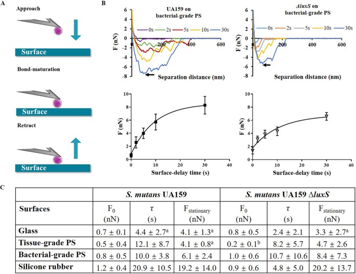
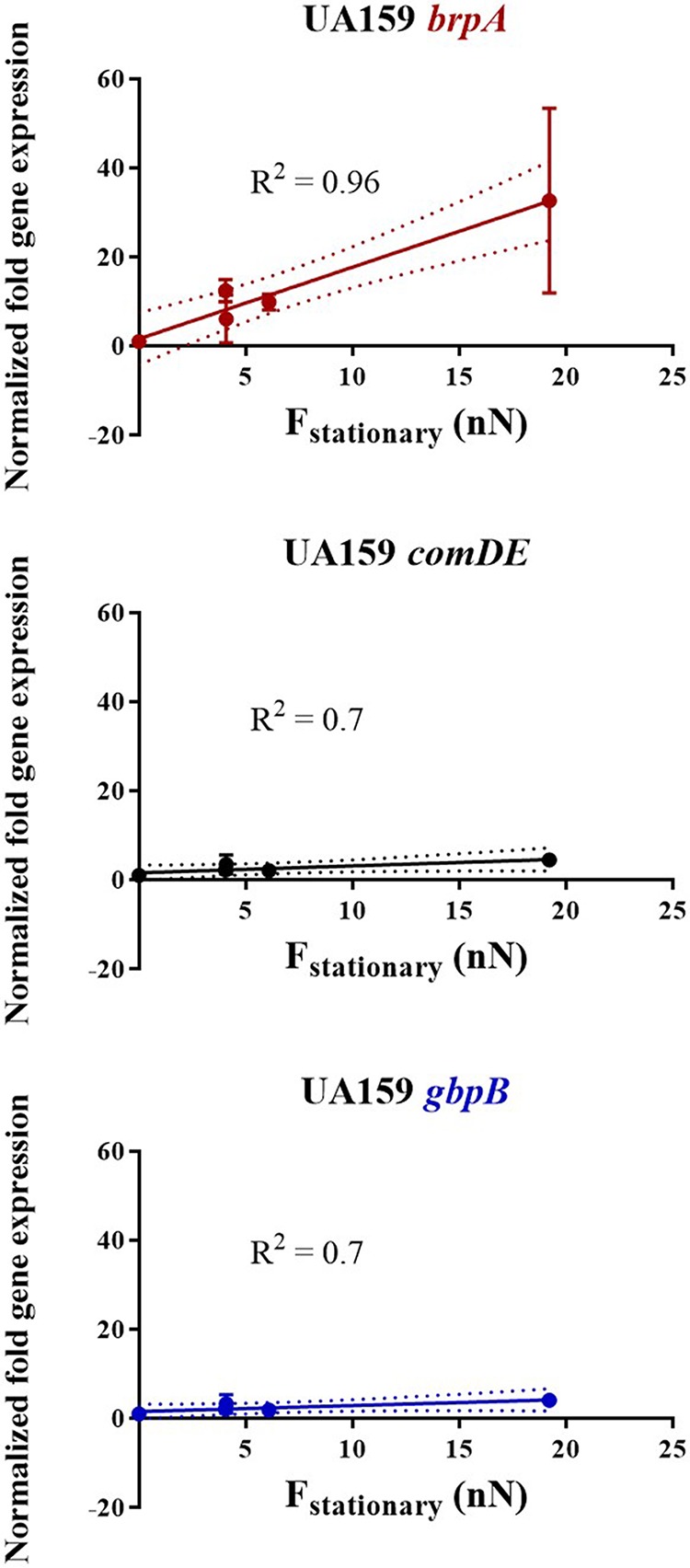
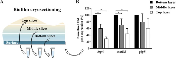
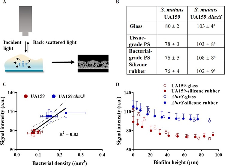
Bacterial adhesion is accompanied by altered gene expression, leading to "emergent" properties of biofilm bacteria that are alien to planktonic ones. With the aim of revealing the role of environmental adhesion forces in emergent biofilm properties, genes in Streptococcus mutans UA159 and a quorum-sensing-deficient mutant were identified that become expressed after adhesion to substratum surfaces. Using atomic force microscopy, adhesion forces of initial S. mutans colonizers on four different substrata were determined and related to gene expression. Adhesion forces upon initial contact were similarly low across different substrata, ranging between 0.2 and 1.2 nN regardless of the strain considered. Bond maturation required up to 21 s, depending on the strain and substratum surface involved, but stationary adhesion forces also were similar in the parent and in the mutant strain. However, stationary adhesion forces were largest on hydrophobic silicone rubber (19 to 20 nN), while being smallest on hydrophilic glass (3 to 4 nN). brpA gene expression in thin (34 to 48 μm) 5-h S. mutans UA159 biofilms was most sensitive to adhesion forces, while expression of gbpB and comDE expressions was weakly sensitive. ftf, gtfB, vicR, and relA expression was insensitive to adhesion forces. In thicker (98 to 151 μm) 24-h biofilms, adhesion-force-induced gene expression and emergent extracellular polymeric substance (EPS) production were limited to the first 20 to 30 μm above a substratum surface. In the quorum-sensing-deficient S. mutans, adhesion-force-controlled gene expression was absent in both 5- and 24-h biofilms. Thus, initial colonizers of substratum surfaces sense adhesion forces that externally trigger emergent biofilm properties over a limited distance above a substratum surface through quorum sensing.IMPORTANCE A new concept in biofilm science is introduced: "adhesion force sensitivity of genes," defining the degree up to which expression of different genes in adhering bacteria is controlled by the environmental adhesion forces they experience. Analysis of gene expression as a function of height in a biofilm showed that the information about the substratum surface to which initially adhering bacteria adhere is passed up to a biofilm height of 20 to 30 μm above a substratum surface, highlighting the importance and limitations of cell-to-cell communication in a biofilm. Bacteria in a biofilm mode of growth, as opposed to planktonic growth, are responsible for the great majority of human infections, predicted to become the number one cause of death in 2050. The concept of adhesion force sensitivity of genes provides better understanding of bacterial adaptation in biofilms, direly needed for the design of improved therapeutic measures that evade the recalcitrance of biofilm bacteria to antimicrobials.

Keywords: OCT; atomic force microscopy; quorum sensing; regulation of gene expression; surface sensing.


Figures

FIG 1
FIG 1
Bacterial adhesion force characteristics of both streptococcal strains on four substratum surfaces with different hydrophobicities. (A) Schematics of single-bacterial-contact probe atomic force microscopy. A bacterium is attached to a tipless AFM cantilever and brought to contact with a substratum surface, after which the cantilever is retracted following a surface delay that can be varied up to a maximum of 30 s. Upon retraction, the adhesion force by which the bacterium was attracted to the surface can be calculated from the cantilever bending. (B) Example of retraction force-distance curves taken after different surface delay times for S. mutans UA159 on a bacterial-grade polystyrene (PS) surface. (The arrow points to the force value, taken as the adhesion force.) Also included is a graph of streptococcal adhesion forces as a function of surface delay time for the parent strain and its quorum-sensing-deficient isogenic mutant. (C) Initial and stationary streptococcal adhesion forces F0 and Fstationary, together with the characteristic bond maturation time constant τ on the different substratum surfaces. All data represent averages over 8 spots on 4 different surfaces of each substratum, measured with 4 different probes and bacteria from 4 different cultures, with ± signs representing standard deviation (SD) values over 32 measurements. Superscript letters in panel C indicate statistical significance as follows: a, statistically significant (P < 0.05, one-way ANOVA) differences from silicone rubber; b, statistically significant (P < 0.05, one-way ANOVA) differences between tissue-grade and bacterial-grade PS surfaces.
FIG 2
FIG 2
Normalized fold gene expression with significant relationships to adhesion forces in S. mutans UA159 as a function of the stationary adhesion force to different substratum surfaces over the entire height of 5-h biofilms. Error bars denote SD values in fold gene expression over triplicate experiments, while the solid lines represent assumed linear relationships through the data points, with the correlation coefficient R2 as presented. Dotted lines represent 95% confidence intervals.
FIG 3
FIG 3
Gene expression in different layers of 24-h S. mutans UA159 biofilm on a silicone rubber surface. (A) Schematics of biofilm cryosectioning and gene expression in three biofilm slices taken at different heights in the biofilm above the substratum surface. (B) Percentage of normalized (with respect to the internal 16S rRNA control) adhesion-force-induced expression of selected genes at different heights above a silicone rubber surface in 24-h S. mutans UA159 biofilm, expressed relative to gene expression in the bottom layer of the biofilm closest to the substratum surface, set at 100%. Error bars denote SD values over triplicate experiments. *, statistically different at P < 0.05 by one-way ANOVA.
FIG 4
FIG 4
Analysis of OCT images of 24-h S. mutans UA159 and UA159 ΔluxS biofilms. (A) Schematics of signal intensity development by back-scattered light in OCT: based on an artificial whiteness scale, bacteria yield white regions (high signal intensity) due to back-scattering, while water- and soluble EPS-filled pockets do not back-scatter light and appear as black regions (low signal intensity). (B) Average signal intensity over an entire biofilm in 24-h streptococcal biofilms on the four different substratum surfaces. The superscript letter a in panel B indicates significant difference between S. mutans UA159 and UA159 ΔluxS (P < 0.05, Mann-Whitney test). (C) Average signal intensity over an entire biofilm as a function of the volumetric bacterial density for 24-h streptococcal biofilms of both strains on the four different substratum surfaces. Dotted lines represent 95% confidence intervals. (D) Local signal intensity in OCT images of 24-h streptococcal biofilms on glass and silicone rubber as a function of the biofilm height above the substratum surface. There are no statistically significant (P > 0.05, Mann-Whitney test) differences at corresponding heights for the mutant strain on hydrophobic silicone rubber and hydrophilic glass, while for the parent strain, signal intensities are lower on silicone rubber than on hydrophilic glass up to a thickness of 20 to 25 μm. Error bars indicate SD over different experiments with separately cultured bacteria (n = 3).

Similar articles

Cited by

References

    1. Tolker-Nielsen T. 2015. Biofilm development. Microbiol Spectr doi:10.1128/microbiolspec.MB-0001-2014. - DOI - PubMed
    1. Hermansson M. 1999. The DLVO theory in microbial adhesion. Colloids Surf B Biointerfaces 14:105–119. doi:10.1016/S0927-7765(99)00029-6. - DOI
    1. Carniello V, Peterson BW, van der Mei HC, Busscher HJ. 2018. Physico-chemistry from initial bacterial adhesion to surface-programmed biofilm growth. Adv Colloid Interface Sci 261:1–14. doi:10.1016/j.cis.2018.10.005. - DOI - PubMed
    1. Ren Y, Wang C, Chen Z, Allan E, van der Mei HC, Busscher HJ. 2018. Emergent heterogeneous microenvironments in biofilms: substratum surface heterogeneity and bacterial adhesion force-sensing. FEMS Microbiol Rev 42:259–272. doi:10.1093/femsre/fuy001. - DOI - PubMed
    1. Harapanahalli AK, Younes JA, Allan E, van der Mei HC, Busscher HJ. 2015. Chemical signals and mechanosensing in bacterial responses to their environment. PLoS Pathog 11:e1005057. doi:10.1371/journal.ppat.1005057. - DOI - PMC - PubMed

Publication types

MeSH terms

LinkOut - more resources