. 2019 Nov 5;9(11):3555-3565.
doi: 10.1534/g3.119.400704.

Pharmaco-Genetic Screen To Uncover Actin Regulators Targeted by Prostaglandins During Drosophila Oogenesis

Affiliations

Pharmaco-Genetic Screen To Uncover Actin Regulators Targeted by Prostaglandins During Drosophila Oogenesis

Andrew J Spracklen et al. G3 (Bethesda). .

Abstract

Prostaglandins (PGs) are lipid signaling molecules with numerous physiologic functions, including pain/inflammation, fertility, and cancer. PGs are produced downstream of cyclooxygenase (COX) enzymes, the targets of non-steroidal anti-inflammatory drugs (NSAIDs). In numerous systems, PGs regulate actin cytoskeletal remodeling, however, their mechanisms of action remain largely unknown. To address this deficiency, we undertook a pharmaco-genetic interaction screen during late-stage Drosophila oogenesis. Drosophila oogenesis is as an established model for studying both actin dynamics and PGs. Indeed, during Stage 10B, cage-like arrays of actin bundles surround each nurse cell nucleus, and during Stage 11, the cortical actin contracts, squeezing the cytoplasmic contents into the oocyte. Both of these cytoskeletal properties are required for follicle development and fertility, and are regulated by PGs. Here we describe a pharmaco-genetic interaction screen that takes advantage of the fact that Stage 10B follicles will mature in culture and COX inhibitors, such as aspirin, block this in vitro follicle maturation. In the screen, aspirin was used at a concentration that blocks 50% of the wild-type follicles from maturing in culture. By combining this aspirin treatment with heterozygosity for mutations in actin regulators, we quantitatively identified enhancers and suppressors of COX inhibition. Here we present the screen results and initial follow-up studies on three strong enhancers - Enabled, Capping protein, and non-muscle Myosin II Regulatory Light Chain. Overall, these studies provide new insight into how PGs regulate both actin bundle formation and cellular contraction, properties that are not only essential for development, but are misregulated in disease.

Keywords: Capping protein; Drosophila melanogaster; Enabled; non-muscle myosin II regulatory light chain; prostaglandins.


Figures

Figure 1
Figure 1
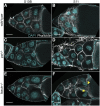
Pxt is required for actin remodeling during S10B and cellular contraction during S11. A-F. Maximum projection images of 2-4 confocal slices of S10B and S11 follicles of the indicated genotypes. Stage was determined in the mutants by centripetal follicle cell migration. DAPI (cyan) and phalloidin (white). Scale bars = 50μm. A-B. wild-type. C-D pxtf/f. E-F. fascinsn28/sn28. Yellow arrowheads indicate nurse cell nuclei that have plugged the ring canals during dumping. Loss of Pxt results in failure of nurse cell dumping; follicles fail to properly form actin bundles (C) in the nurse cells and don’t undergo nurse cell contraction (D). Conversely, while loss of Fascin results in a lack of actin bundles in the nurse cells (E), follicles still undergo contraction as evident by nurse cell nuclei plugging the ring canals (F, yellow arrowheads).
Figure 2
Figure 2
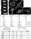
In vitro egg maturation examples and screen rationale. A. Representative images of S10B-S14 Drosophila follicles taken using a stereo dissecting scope; anterior is at the left. Blue asterisks indicate the oocyte, yellow brackets mark the nurse cells, and the white arrow indicates the dorsal appendages in S14. In the IVEM assay, follicles that remain in S10B-11 are scored as having failed to dump, whereas follicles that have reached S12-14 are scored as having completed dumping. B-F. Representative images of in vitro maturing follicles at the start (B) or end of the experiment (C-F) in control media (B-C, vehicle) or aspirin media (D-F, IC50 aspirin). Follicles failing to dump are marked with red asterisks. G. Diagrams illustrating the rationale behind the pharmaco-genetic interaction screen. H. Formulas for calculating the dumping index and normalized dumping index (the colors match the data in the table in I). I. Table of the data and example calculations from two experiments – one for an enhancer (ena210) and one for a suppressor (shgE17B). In control media, the majority of wild-type follicles complete nurse cell dumping (C, 92%), while in aspirin media only 50% complete dumping (D, G). The dumping index for control follicles is expected to be around 0.5 (H, I). A genetic enhancer will result in <50% completing dumping in aspirin media (E [23%] and G), a dumping index of <0.5, and a negative normalized dumping index (I, ena210). A genetic suppressor will result in >50% completing dumping (F [73%] and G), a dumping index of >0.5, and a positive normalized dumping index (I, shgE17B). Reduced levels of an actin-binding protein that is not a downstream target of PG signaling during S10B would not be expected to modify follicle sensitivity to COX inhibition, resulting in 50% completing dumping (G), a dumping index of ∼0.5, and a normalized dumping index of ∼0. Scale bars = 50μm.
Figure 3
Figure 3
Pharmaco-genetic interaction screen reveals multiple actin- binding proteins as candidate downstream targets of prostaglandin signaling. Chart of the normalized dumping indices for all genotypes tested in the pharmaco-genetic interaction screen. Wild-type control values were set to zero and normalized experimental dumping indices were calculated by subtracting the dumping index of the experimental group from the wild-type dumping index for each individual experiment (see Figure 2H). Dashed red lines indicate ± 1 SD (±0.054) from all wild-type control values. Solid red lines represent ± 3 SD (±0.162) from all wild-type control values. Genotypes exhibiting normalized dumping indices between 0 and ± 1 SD from wild-type control values are classified as non-interactors. Genotypes exhibiting normalized dumping indices falling between ±1 to ± 3 SD from wild-type control values are classified as weak interactors. Genotypes exhibiting normalized dumping indices falling outside of ±3 SD from wild-type control values are classified as strong interactors. Error bars = standard deviation (SD).
Figure 4
Figure 4
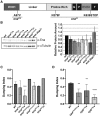
Altering Ena activity strongly sensitizes S10B follicles to the effects of COX inhibition. A. Schematic detailing Ena protein structure and the mutations carried by the ena23 and ena210 alleles. EVH = Ena/VASP homology domain; G= G-actin binding domain, F= F-actin binding domain, T= tetramerization domain. B. Representative western blot and quantification of Ena levels for genotypes indicated. C. Graph of pharmaco-genetic interaction data for ena alleles. D. Graph of pharmaco-interaction data for cpa and cpb alleles. Heterozygosity for ena23, ena210, enaGC1, enaGC5, or enaDG14706 or homozygosity for enaEY13131 results in a mild decrease in total Ena levels compared to wild-type controls (B, n = 2, error bars = SD; note both full-length and truncated Ena are observed in the ena23/+ lane). Additionally, heterozygosity for an Ena duplication (ena dup; Dp(2;2)Cam18/+) results in a mild increase in total Ena levels compared to wild-type controls (B). Heterozygosity for enaGC1, enaGC5, enaDG14706, and ena23, as well as homozygosity enaEY13131, fail to modify follicle sensitivity to the effects of COX inhibition (C). However, heterozygosity for the ena210 allele significantly enhances the ability of 1.5mM aspirin to inhibit nurse cell dumping compared to wild-type controls (C). Heterozygosity the alpha-subunit of Capping protein (cpaKG02261), and the beta-subunit of Capping protein (cpb6.15 and cpb19F) significantly enhance the ability of 1.5mM aspirin to block nurse cell dumping compared to wild-type controls (D). The number of trials (n) are indicated in Table S1. Error bars = SD. **** P < 0.0001, *** P < 0.001, **P < 0.01, and *P < 0.05.
Figure 5
Figure 5
Prostaglandins pharmaco-genetically interact with and regulate the activity of MRLC. A. Graph of pharmaco-interaction data for sqh alleles; sqh encodes the Drosophila MRLC. B. Representative western blot and quantification of Zipper (Drosophila MHC) levels for genotypes indicated. Tubulin was used as a loading control and Zipper protein levels were normalized to Tubulin. C-D”. Maximum projections of 2-3 confocal slices of S10 follicles of the indicated genotypes. C, D. Phosphorylated myosin regulatory light chain (S19), white. C’, D’. Phosphorylated myosin regulatory light chain (S19) pseudocolored with Rainbow RGB, red indicating the highest intensity pixels. Insets are zoom-ins of the nurse cell membranes within the yellow squares. C’’, D’’. Phalloidin (F-actin), white. E. Graph showing quantification of p-MRLC intensity in arbitrary units (AU) along the membrane of a posterior nurse cell of a S10B follicle of the indicated genotypes. Briefly, the average fluorescent intensity of p-MRLC was measured for the same length along a posterior nurse cell membrane for each of the indicated genotypes; each circle represents a single follicle. Heterozygosity for sqhAX3 or sqhEY09875 enhances the ability of aspirin to inhibit nurse cell dumping compared to wild-type controls, and heterozygosity for sqhDf(1)5D results in a mild increase in aspirin’s ability to inhibit nurse cell dumping (A). Loss of PG signaling using two different pxt alleles, pxtEY/EY or pxtf/f, does not alter total Zipper (Drosophila MHC) levels (B; n = 3, error bars = SD). Loss of PG signaling alters phospho-MRLC localization on nurse cell membranes with the pxt mutants exhibiting patchy localization throughout the follicle and aberrant enrichment on the anterior nurse cells (C-D’). The change in phospho-MRLC is not due to cortical actin breakdown, as the cortical actin is intact in the pxt mutant (C’’, D’’). Loss of Pxt results in reduced activated MRLC along the posterior nurse cell membranes (E). Error bars = SD. ns = P > 0.05 and **** P < 0.0001. Scale bars= 50 μm.

Similar articles

Cited by

References

    1. Abramoff M. D., Magalhaes P. J., and Ram S. J., 2004. Image Processing with ImageJ. Biophoton. Int. 11: 36–42.
    1. Ahern-Djamali S. M., Comer A. R., Bachmann C., Kastenmeier A. S., Reddy S. K. et al. , 1998. Mutations in Drosophila enabled and rescue by human vasodilator-stimulated phosphoprotein (VASP) indicate important functional roles for Ena/VASP homology domain 1 (EVH1) and EVH2 domains. Mol. Biol. Cell 9: 2157–2171. 10.1091/mbc.9.8.2157 - DOI - PMC - PubMed
    1. Allaj V., Guo C., and Nie D., 2013. Non-steroid anti-inflammatory drugs, prostaglandins, and cancer. Cell Biosci. 3: 8 10.1186/2045-3701-3-8 - DOI - PMC - PubMed
    1. Amano M., Ito M., Kimura K., Fukata Y., Chihara K. et al. , 1996. Phosphorylation and activation of myosin by Rho-associated kinase (Rho-kinase). J. Biol. Chem. 271: 20246–20249. 10.1074/jbc.271.34.20246 - DOI - PubMed
    1. Aranjuez G., Burtscher A., Sawant K., Majumder P., and McDonald J. A., 2016. Dynamic myosin activation promotes collective morphology and migration by locally balancing oppositional forces from surrounding tissue. Mol. Biol. Cell 27: 1898–1910. 10.1091/mbc.e15-10-0744 - DOI - PMC - PubMed

Publication types

MeSH terms

LinkOut - more resources