. 2019 Nov;213(3):923-939.
doi: 10.1534/genetics.119.302670. Epub 2019 Sep 10.

Germline Maintenance Through the Multifaceted Activities of GLH/Vasa in Caenorhabditis elegans P Granules

Affiliations

Germline Maintenance Through the Multifaceted Activities of GLH/Vasa in Caenorhabditis elegans P Granules

Elisabeth A Marnik et al. Genetics. 2019 Nov.

Abstract

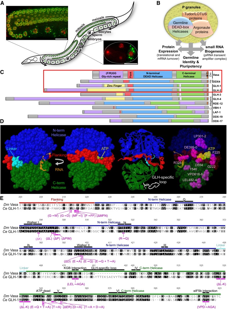
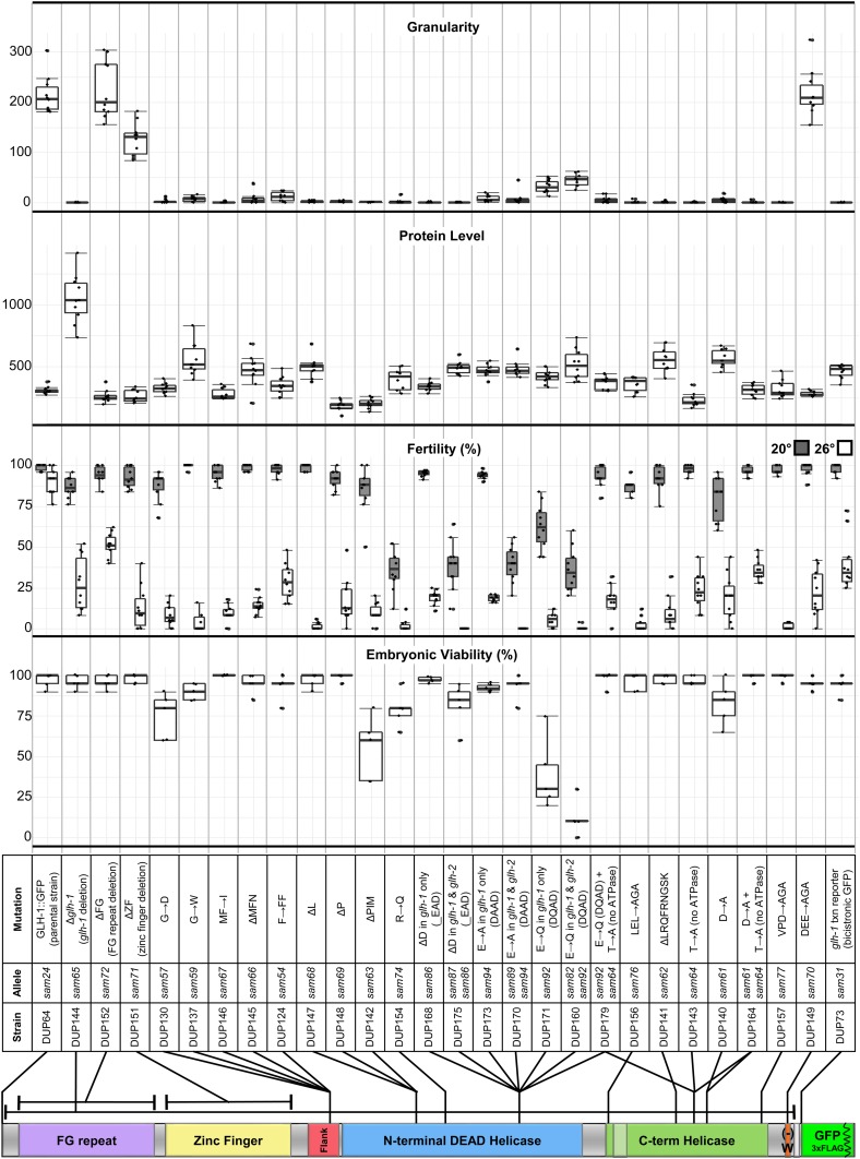
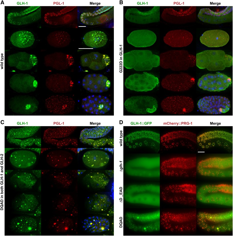
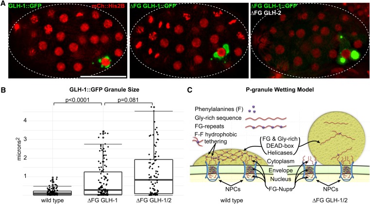
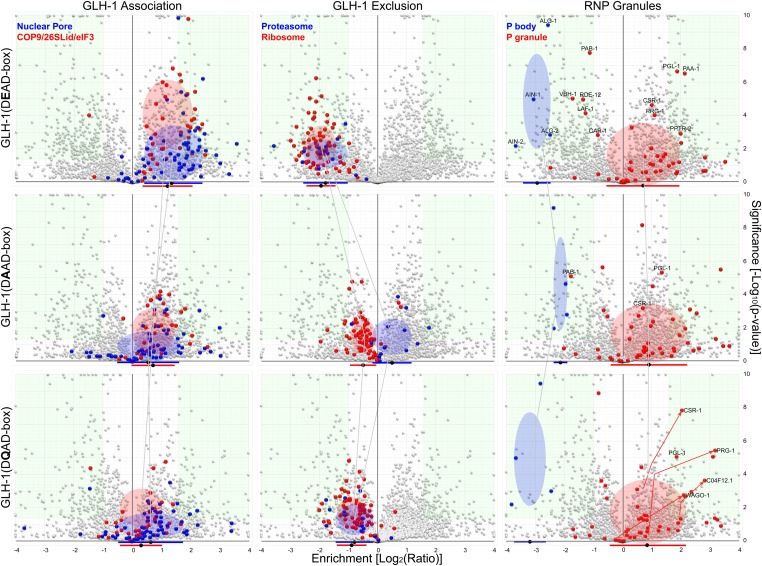
Vasa homologs are ATP-dependent DEAD-box helicases, multipotency factors, and critical components that specify and protect the germline. They regulate translation, amplify piwi-interacting RNAs (piRNAs), and act as RNA solvents; however, the limited availability of mutagenesis-derived alleles and their wide range of phenotypes have complicated their analysis. Now, with clustered regularly interspaced short palindromic repeats (CRISPR/Cas9), these limitations can be mitigated to determine why protein domains have been lost or retained throughout evolution. Here, we define the functional motifs of GLH-1/Vasa in Caenorhabditis elegans using 28 endogenous, mutant alleles. We show that GLH-1's helicase activity is required to retain its association with P granules. GLH-1 remains in P granules when changes are made outside of the helicase and flanking domains, but fertility is still compromised. Removal of the glycine-rich repeats from GLH proteins progressively diminishes P-granule wetting-like interactions at the nuclear periphery. Mass spectrometry of GLH-1-associated proteins implies conservation of a transient piRNA-amplifying complex, and reveals a novel affinity between GLH-1 and three structurally conserved PCI (26S Proteasome Lid, COP9, and eIF3) complexes or "zomes," along with a reciprocal aversion for assembled ribosomes and the 26S proteasome. These results suggest that P granules compartmentalize the cytoplasm to exclude large protein assemblies, effectively shielding associated transcripts from translation and associated proteins from turnover. Within germ granules, Vasa homologs may act as solvents, ensuring mRNA accessibility by small RNA surveillance and amplification pathways, and facilitating mRNA export through germ granules to initiate translation.

Keywords: C. elegans; DDX4; GLH-1; P granules; PCI complex; Vasa; germ granules; germline.


Figures

Figure 1
Figure 1
GLH Proteins in the C. elegans germline. (A) GLH-1::GFP::3xFLAG in P granules and mCherry:His2B-marked chromatin. Inserts provide context for expression in the germline loop and four-cell embryo. (B) Schematic depicting the function of core proteins in P granules. (C) Conservation of Vasa/DDX4-like DEAD-box helicases in C. elegans. A red box surrounds proteins that contain all four Vasa-defining domains (glycine-rich in purple, flanking in red, N- and C- terminal helicase in blue and green, and negatively charged residues before a terminal tryptophan in orange). The GLH-specific loop is shown in white. (D) Crystal structure of Vasa showing front and back views of the flanking and helicase domains, in relation to ATP- and RNA-binding pockets [as determined by Sengoku et al. (2006)]. Image 3 is an overlay of Vasa (ribbon) with an iTasser-predicted model of GLH-1 (backbone) that shows the location of the GLH-specific loop (white). Image 4 shows key amino acid residues targeted in this study and their location within the Vasa protein structure. (E) Sequence alignment of the flanking and helicase domains in Drosophila Vasa with C. elegans GLH-1. Protein domains and mutations are indicated (purple). The K295A mutation was not obtained. The ΔER550-1 was not sustainable. See also Figure S1. Ce, C. elegans; ; Dm, Drosophila melanogaster; Hs, Homo sapiens; piRNA, piwi-interacting RNA.
Figure 2
Figure 2
Level and distribution of mutant forms of GLH-1 in C. elegans. (A) GLH-1::GFP::3xFLAG expression in the germline loop. Fixed exposures (left) were normalized (middle) to better view the distribution of fluorescence. GLH-1 granules were quantified using ImageJ (right). In ΔPIM and DQAD mutants, embryos arrest in the elongation phase. (C) GLH-1(DQAD)::GFP::3xFLAG accumulation in proximal (left) and distal (right) germlines is enhanced in GLH-2(DQAD). In double mutants, GLH-1(DQAD) aggregates persist in somatic blastomeres and the soma of hatched worms (bottom). Quantification was performed on images from 10 worms for each strain (see Figure S2). PGC, primordial germ cell.
Figure 3
Figure 3
Consequences of GLH-1 mutant alleles. From top to bottom: comparison of granularity (GLH-1 granules), GLH-1 protein level, fertility at permissive (20°) and restrictive (26°) temperatures, and embryonic viability in GLH-1 mutants. Mutation details, strains, and allele names and their respective locations are indicated. Replicates for each of the four assays are provided in the Materials and Methods. Box plots represent quartiles above and below the median with whiskers extending 1 SD from the mean. See also Figure S2. C-term, C-terminal.
Figure 4
Figure 4
Colocalization of P-granule components in wild-type and GLH-1 mutants. (A–C) Immunostaining of GLH-1 (green) and PGL-1 (red) in fixed germlines, and 1-, 4-, 16-, and 32-cell embryos. (D) GLH-1::GFP and mCherry::PRG-1 in the germline of living worms. At least 10 worms were imaged for each indicated genotype.
Figure 5
Figure 5
Glycine-rich FG repeats tether GLH-1 to the nuclear periphery. (A) Distribution of GLH-1::GFP (green) in the germline blastomere P4 of a wild-type embryo (left), an embryo lacking FG repeats in GLH-1 (middle), and an embryo lacking FG repeats in both GLH-1 and GLH-2 (right). mCherry::H2B (red) marks chromatin. (B) GLH-1-granule sizes in the germline blastomere P4 quantified using ImageJ. (C) Model depicting the hydrophobic tethering of P-granule FG-repeat domains to FG-nucleoporins (Nups) at the nuclear periphery. For size assays, P granules were counted from ≥ 40 embryos for each strain. Box plots show median quartiles and 1 SD above the mean. NPC, nuclear pore complex.
Figure 6
Figure 6
GLH-1 protein associations. Volcano plots show the significance and enrichment of proteins that immunoprecipitate with GLH-1::GFP::3xFLAG over the glh-1 transcriptional reporter expressing GFP::3xFLAG alone, as identified by LC-MS/MS. Left column: protein families showing an enriched GLH-1 association include nuclear pore proteins (blue) and subunits of PCI scaffolding complexes. These associations decrease in DAAD (middle row) and DQAD (bottom row) mutants. Red and blue bars under the x-axis indicate the median and 1 SD, and colored ovals do the same but also indicate the distribution of significance. Green boxes show normalized enrichment and exclusion > 2.5-fold, and a P-value < 0.05. Nuclear pore genes used in this analysis are indicated in Table S3. Middle column: protein families showing stronger enrichment for GFP::3xFLAG alone include 20S core subunits of the 26S proteasome (blue) and subunits of the ribosome (red). Right column: proteins associated with RNP granules. P-body proteins (blue) and P-granule proteins (red). Data were obtained from two technical replicates for each indicated genotype. See also Table S3. LC, liquid chromatography; MS, mass spectrometry; PCI, 26S Proteasome Lid, COP9 Signalosome, and eIF3; RNP, ribonucleoproteins.
Figure 7
Figure 7
Ribosomes and the size exclusion properties of P granules. (A) Immunostaining of RPL-7a (red) with GLH-1::GFP. RPL-7a is more concentrated in the shared cytoplasm of the rachis (arrows) than germ granule-rich areas at the perimeter (arrowheads), but it is not excluded from P granules. (B) 18S rRNA FISH signal (red) is saturated in the cytoplasm of germ cells where it associates with the 40S ribosomal subunit, but is excluded from GLH-1::GFP-marked P (arrows) and yolk granules (arrowheads).
Figure 8
Figure 8
Summary figure. Top left; GLH-1 represented as an oval with its N-terminal (blue), flanking (red), and C-terminal helicase domains. GLH-1’s association in P granules (yellow outline) requires cycles of RNA and adenosine triphosphate (ATP) binding (closed) and release of adenosine diphosphate (ADP) and phosphate (Pi). Top right; GLH-1 is dispersed in mutants that inhibit substrate binding (_EAD, DAAD) or substrate release (DQAD), and in the latter case GLH-1(DQAD) forms aggregates with specific Argonaute (AGO) proteins. Bottom left; GLH-1 associates with the PCI complexes (26S Proteasome Lid, COP9, and eIF3) while excluding ribosomal subunits (60S and 40S) and the 26S proteasome. Bottom right; The phenylalanine - glycine (FG) repeats of GLH-1 tether P granules to FG-nucleoporin proteins.

References

    1. Andralojc K. M., Campbell A. C., Kelly A. L., Terrey M., Tanner P. C. et al. , 2017. ELLI-1, a novel germline protein, modulates RNAi activity and P-granule accumulation in Caenorhabditis elegans. PLoS Genet. 13: e1006611 10.1371/journal.pgen.1006611 - DOI - PMC - PubMed
    1. Brenner S., 1974. The genetics of Caenorhabditis elegans. Genetics 77: 71–94. - PMC - PubMed
    1. Campbell A. C., and Updike D. L., 2015. CSR-1 and P granules suppress sperm-specific transcription in the C. elegans germline. Development 142: 1745–1755. 10.1242/dev.121434 - DOI - PMC - PubMed
    1. Carrera P., Johnstone O., Nakamura A., Casanova J., Jäckle H. et al. , 2000. VASA mediates translation through interaction with a Drosophila yIF2 homolog. Mol. Cell 5: 181–187. 10.1016/S1097-2765(00)80414-1 - DOI - PubMed
    1. Cruciat C.-M., Dolde C., de Groot R. E. A., Ohkawara B., Reinhard C. et al. , 2013. RNA helicase DDX3 is a regulatory subunit of casein kinase 1 in Wnt-β-catenin signaling. Science 339: 1436–1441. 10.1126/science.1231499 - DOI - PubMed

Publication types

MeSH terms