. 2019 Sep 24;116(39):19619-19625.
doi: 10.1073/pnas.1908706116. Epub 2019 Sep 10.

The single-cell transcriptomic landscape of early human diabetic nephropathy

Affiliations

The single-cell transcriptomic landscape of early human diabetic nephropathy

Parker C Wilson et al. Proc Natl Acad Sci U S A. .

Abstract

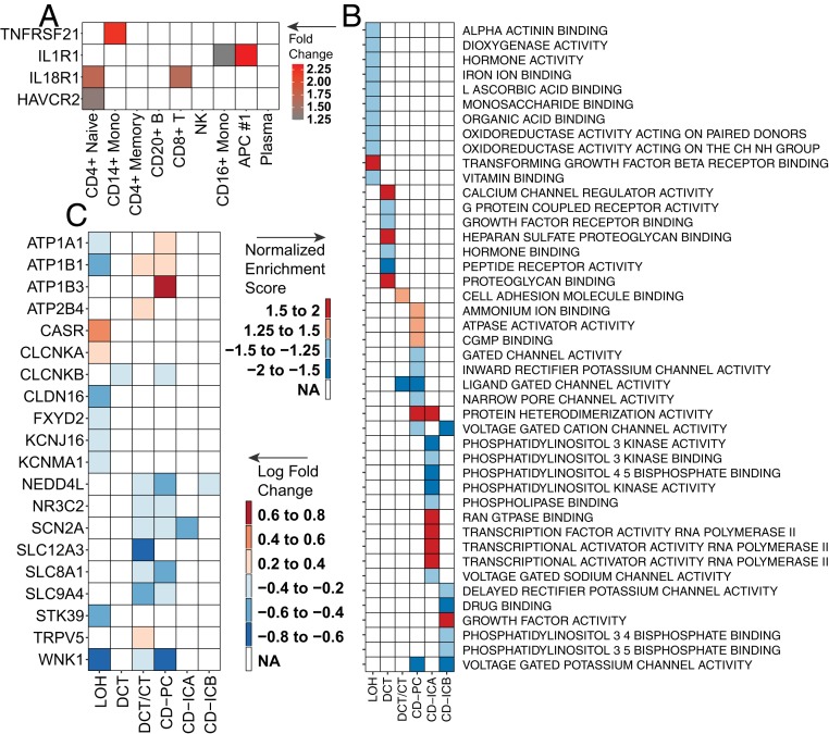
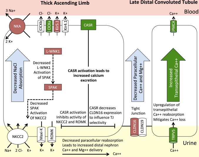
Diabetic nephropathy is characterized by damage to both the glomerulus and tubulointerstitium, but relatively little is known about accompanying cell-specific changes in gene expression. We performed unbiased single-nucleus RNA sequencing (snRNA-seq) on cryopreserved human diabetic kidney samples to generate 23,980 single-nucleus transcriptomes from 3 control and 3 early diabetic nephropathy samples. All major cell types of the kidney were represented in the final dataset. Side-by-side comparison demonstrated cell-type-specific changes in gene expression that are important for ion transport, angiogenesis, and immune cell activation. In particular, we show that the diabetic thick ascending limb, late distal convoluted tubule, and principal cells all adopt a gene expression signature consistent with increased potassium secretion, including alterations in Na+/K+-ATPase, WNK1, mineralocorticoid receptor, and NEDD4L expression, as well as decreased paracellular calcium and magnesium reabsorption. We also identify strong angiogenic signatures in glomerular cell types, proximal convoluted tubule, distal convoluted tubule, and principal cells. Taken together, these results suggest that increased potassium secretion and angiogenic signaling represent early kidney responses in human diabetic nephropathy.

Keywords: RNA-seq; diabetic nephropathy; single cell.


Conflict of interest statement

Conflict of interest statement: In separate work, B.D.H. consults for and receives grant support from Janssen to study mouse diabetic nephropathy. B.D.H. also consults for and holds equity in Chinook Therapeutics. Neither relationship is related to the contents of this manuscript.

Figures

Fig. 1.
Fig. 1.
Integrated snRNA-seq dataset of diabetic and control samples. (A) Diabetic and control samples were integrated into a single dataset and clustered using Seurat. PCT, proximal convoluted tubule; CFH, complement factor H; LOH, loop of Henle; DCT, distal convoluted tubule; CT, connecting tubule; CD, collecting duct; PC, principal cell; IC, intercalated cell; PODO, podocyte; ENDO, endothelium; MES, mesangial cell; LEUK, leukocyte. (B) Cell clusters were identified by kidney cell lineage-specific marker expression. (C) The leukocyte cluster (LEUK) was extracted from the integrated dataset and subclustered into leukocyte subsets. (D) Leukocyte subsets were identified by expression of lineage-specific markers.
Fig. 2.
Fig. 2.
Differential gene expression and intercellular signaling in the diabetic glomerulus. Diabetic and control samples were integrated into a single dataset, and gene expression was compared within cell types using Seurat. Differentially expressed genes are displayed for (A) podocytes, (B) mesangial cells, and (C) endothelial cells. Ligand–receptor interactions were inferred using a publicly available database (Ramilowski et al., ref. 44) and include (D) differentially expressed ligand–receptor intercellular signaling pathways and (E and F) all possible ligand–receptor signaling pathways in the podocyte and mesangium. FDR, false discovery rate.
Fig. 3.
Fig. 3.
Differential expression of predictive biomarkers and ion transport pathways. (A) Leukocyte subsets extracted from the integrated dataset were interrogated for differential expression of a panel of inflammatory markers (KRIS). (B) Cells from the loop of Henle (LOH) to the collecting duct underwent gene set enrichment analysis using the R package fgsea and were mapped to gene ontology terms. (C) Differentially expressed genes involved in ion transport in the distal nephron were identified using Seurat.
Fig. 4.
Fig. 4.
Coordination of the distal nephron to promote potassium secretion. Genes are depicted as up-regulated (green fill), down-regulated (red fill), or no significant change (white fill) relative to control. The loop of Henle showed decreased expression of the Na+/K+-ATPase (NKA) subunits, ATP1A1, ATP1B1, and FXYD2, and the basolateral potassium channel, KCNJ16 (Kir5.1), in addition to decreased expression of WNK1 and its downstream effector, STK39 (SPAK), which regulate activity of the apical Na+-K+-2Cl− cotransporter (NKCC2). Decreased NKA, KCNJ16, and NKCC2 activity in the loop of Henle are expected to impair transcellular sodium and potassium reabsorption. These changes were accompanied by increased expression of NKA and SGK1 with decreased expression of an important regulator of ENaC and potassium secretion, NEDD4L, in the collecting duct.
Fig. 5.
Fig. 5.
Decreased paracellular reabsorption of calcium and magnesium. Genes are depicted as up-regulated (green fill), down-regulated (red fill), or no significant change (white fill) relative to controls. Decreased NKA, KCNJ16 (Kir5.1), and NKCC2 activity in the loop of Henle are expected to impair transcellular sodium and potassium reabsorption and decrease paracellular reabsorption of calcium and magnesium. This would be exacerbated by the observed increased expression of the calcium-sensing receptor (CASR) and decreased expression of CLDN16, which regulates tight junction permeability. Increased expression of the apical calcium-selective channel, TRPV5, and the basolateral plasma membrane calcium ATPase (PMCA) in the late distal convoluted tubule likely result as a compensatory mechanism to prevent excessive urinary calcium loss.

References

    1. Wilson P. C., Humphreys B. D., Single-cell genomics and gene editing: Implications for nephrology. Nat. Rev. Nephrol. 15, 63–64 (2019). - PubMed
    1. Wu H., Kirita Y., Donnelly E. L., Humphreys B. D., Advantages of single-nucleus over single-cell RNA sequencing of adult kidney: Rare cell types and novel cell states revealed in fibrosis. J. Am. Soc. Nephrol. 30, 23–32 (2019). - PMC - PubMed
    1. Colhoun H. M., Marcovecchio M. L., Biomarkers of diabetic kidney disease. Diabetologia 61, 996–1011 (2018). - PMC - PubMed
    1. Lin J. S., Susztak K., Podocytes: The weakest link in diabetic kidney disease? Curr. Diab. Rep. 16, 45 (2016). - PMC - PubMed
    1. Baelde H. J., et al. , Gene expression profiling in glomeruli from human kidneys with diabetic nephropathy. Am. J. Kidney Dis. 43, 636–650 (2004). - PubMed

Publication types

MeSH terms