. 2019 Dec;25(12):1714-1730.
doi: 10.1261/rna.072074.119. Epub 2019 Sep 10.

Circular code motifs in the ribosome: a missing link in the evolution of translation?

Affiliations

Circular code motifs in the ribosome: a missing link in the evolution of translation?

Gopal Dila et al. RNA. 2019 Dec.

Abstract

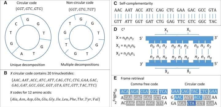
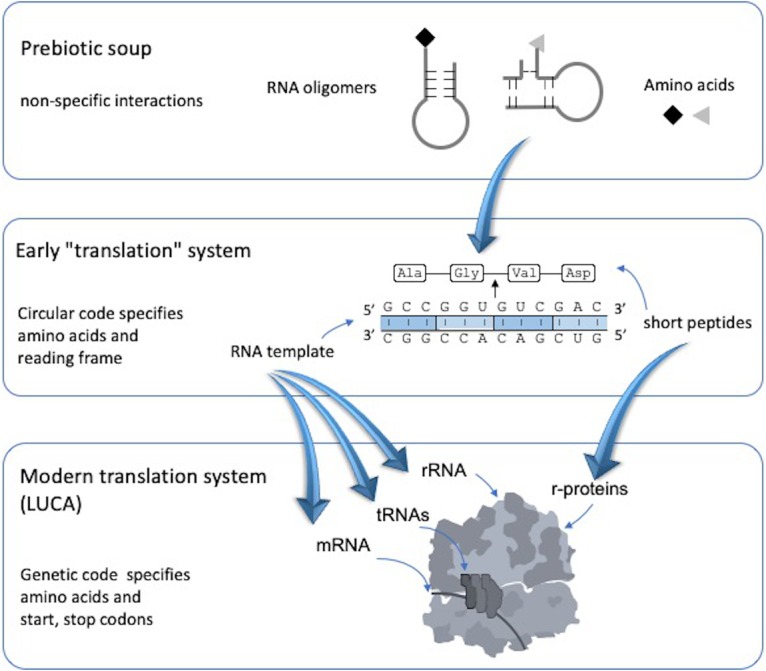
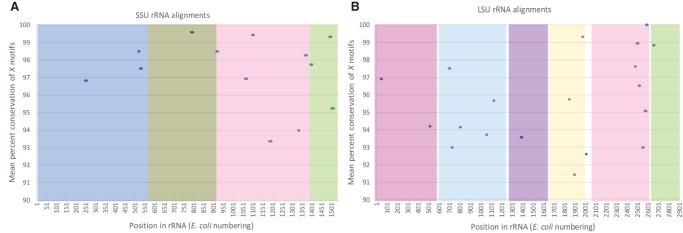
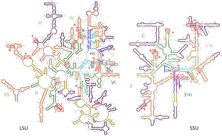
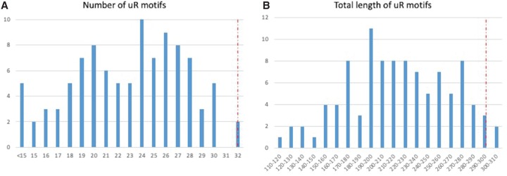
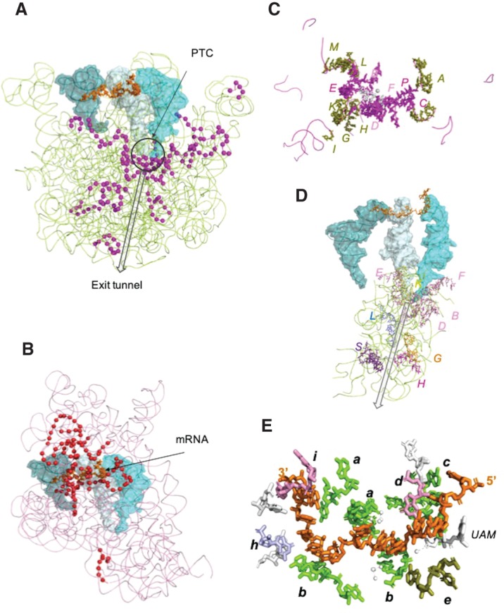
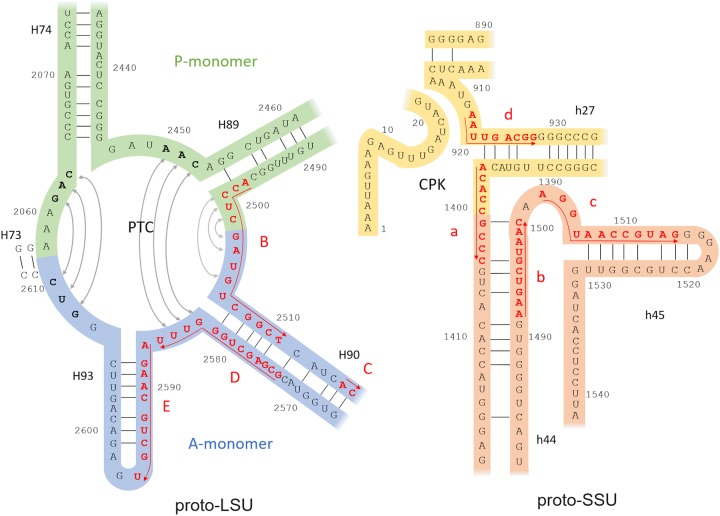
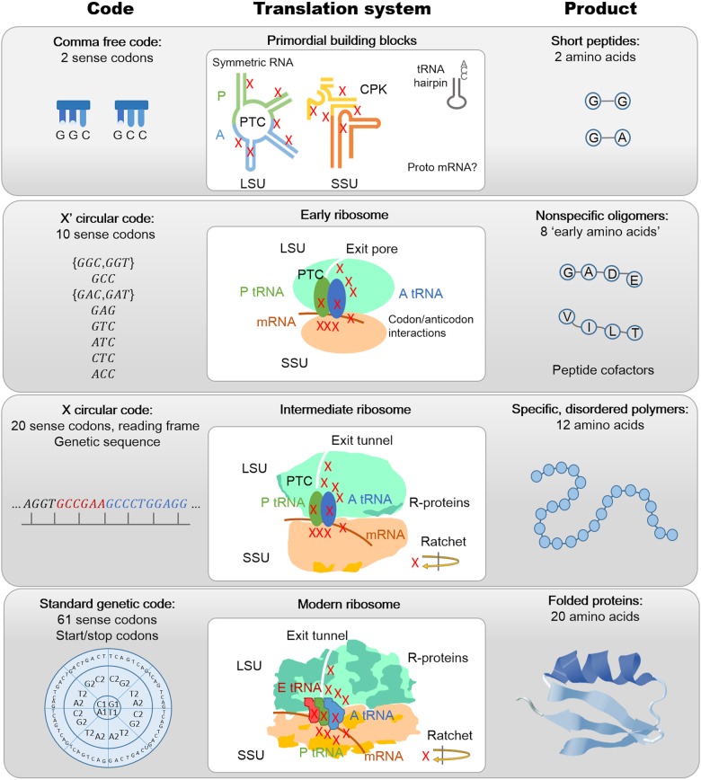
The origin of the genetic code remains enigmatic five decades after it was elucidated, although there is growing evidence that the code coevolved progressively with the ribosome. A number of primordial codes were proposed as ancestors of the modern genetic code, including comma-free codes such as the RRY, RNY, or GNC codes (R = G or A, Y = C or T, N = any nucleotide), and the X circular code, an error-correcting code that also allows identification and maintenance of the reading frame. It was demonstrated previously that motifs of the X circular code are significantly enriched in the protein-coding genes of most organisms, from bacteria to eukaryotes. Here, we show that imprints of this code also exist in the ribosomal RNA (rRNA). In a large-scale study involving 133 organisms representative of the three domains of life, we identified 32 universal X motifs that are conserved in the rRNA of >90% of the organisms. Intriguingly, most of the universal X motifs are located in rRNA regions involved in important ribosome functions, notably in the peptidyl transferase center and the decoding center that form the original "proto-ribosome." Building on the existing accretion models for ribosome evolution, we propose that error-correcting circular codes represented an important step in the emergence of the modern genetic code. Thus, circular codes would have allowed the simultaneous coding of amino acids and synchronization of the reading frame in primitive translation systems, prior to the emergence of more sophisticated start codon recognition and translation initiation mechanisms.

Keywords: circular code; genetic code; origin of life; ribosome evolution; translation.


Figures

FIGURE 1.
FIGURE 1.
Properties of the X circular code. (A) The definition of circularity implies that any word of the X code written on a circle has a unique decomposition. (B) The X circular code is maximal (with 20 trinucleotides) and codes for 12 amino acids. (C) The X code is composed of 10 trinucleotides and their complementary trinucleotides. (D) The permutations of the X code associated with the shifted frames 1 and 2, named X1 and X2, respectively, are circular codes (C3) and in addition are complementary to each other: a word in the shifted frame 1 of the strand 5′–3′ is complementary to the word in the shifted frame 2 of the strand 3′–5′, and vice versa. Note that X1 and X2 are shown in only one strand for simplicity, although they exist in both strands. (E) According to the definition of a comma-free code, all words in the reading frame (frame 0) are valid (shown in blue), whereas all out-of-frame words are invalid (gray). For the X circular code, valid words may be present in frames 1 or 2, up to a length of at most 13 nt.
FIGURE 2.
FIGURE 2.
Hypothesis of circular codes as a missing link in the early evolution of the translation system. The prebiotic soup contained RNA oligomers and amino acids that interacted nonspecifically. They then coevolved to form an ancestral RNA-based “translation” system, with more specific mapping between trinucleotides and amino acids. The RNA template evolved to form the RNA building blocks of the modern ribosome.
FIGURE 3.
FIGURE 3.
(A) Location of the 13 uX motifs in the SSU rRNA alignments (prokaryotic 16S and eukaryotic 18S). The abscissa gives the nucleotide position referenced according to the E. coli 16S rRNA and the ordinate indicates the level of sequence conservation observed in the uX motifs. (B) Location of the 19 uX motifs in the LSU rRNA alignments (prokaryotic 23S and eukaryotic 25/28S). The abscissa gives the nucleotide position referenced according to the Escherichia coli 23S rRNA and the ordinate indicates the level of sequence conservation observed in the uX motifs. Colored boxes indicate rRNA domains (positions in Table 3): for the SSU, light blue for domain 5′, olive for the central domain, pink for 3′M, and green for 3′m domains and for the LSU, magenta for domain I, blue for domain II, violet for domain III, white for domain 0, yellow for domain IV, pink for domain V, and green for domain VI.
FIGURE 4.
FIGURE 4.
Secondary structure schema of the LSU and SSU rRNA (E. coli), showing the location of the uX motifs (red boxes). The schema is colored according to the six phases of the accretion model (Petrov et al. 2015) of ribosome evolution (phase 1, blue; phase 2, cyan; phase 3, green; phase 4, sepia; phase 5, brown; phase 6, purple). uX motifs are labeled with capital letters for LSU motifs and small letters for SSU motifs, according to their order of accretion in the different phases. (PTC) Peptidyl transferase center, (CPK) central pseudoknot.
FIGURE 5.
FIGURE 5.
Distribution of the number and total nucleotide lengths of the uR random motifs in the SSU (16/18S) and LSU (23/28S) rRNA multiple alignments. The corresponding values for the uX motifs are indicated by a vertical red line. (A) Two percent of the random codes have the same number of universal motifs compared to uX motifs (number = 32). (B) Three percent of the random codes have the same or larger total length of universal motifs compared to uX motifs (length = 296).
FIGURE 6.
FIGURE 6.
uX motifs in the rRNA of T. thermophilus. (A) LSU rRNA (green ribbon) with mRNA (orange sticks) and surface representations of tRNAs in the A-site (cyan), P-site (light blue), and E-site (deep teal). Nucleotides of the uX motifs are shown as magenta spheres. The PTC is identified by a black circle and the exit tunnel by a black arrow. (B) SSU rRNA (pink ribbon) with tRNA colored as in A. Nucleotides of the uX motifs are shown as red spheres. (C) Nucleotides in uX motifs close to the PTC (<10 Å in white sticks, <30 Å in magenta sticks, <50 Å in olive sticks). The distances were measured from atom N4 of CYT 2573 (white sphere). All uX motifs are shown as magenta ribbons. (D) All rRNA nucleotides (green ribbons) within 20 Å of the exit tunnel (black arrow) as defined by Dao Duc et al. (2019): nucleotides in uX motifs are colored according to rRNA domains, magenta for domain I, blue for domain II, violet for domain III, orange for domain 0, yellow for domain IV, and pink for domain V (Table 3). tRNA are colored as in A. (E) SSU rRNA nucleotides in contact with mRNA (<5 Å): nucleotides in uX motifs are colored according to rRNA domains, light blue for domain 5′, olive for the central domain, pink for 3′M, and green for 3′m domains (Table 3); other nucleotides and amicoumacin A (UAM) are white. Magnesium ions and their coordinated water molecules are represented by white spheres.
FIGURE 7.
FIGURE 7.
Proto-LSU and proto-SSU, with nucleotides and numbering from the contemporary E. coli 23S and 16S rRNA. uX motifs are highlighted in red and labeled according to the accretion model of Petrov et al. (2015), with 5′–3′ direction indicated by red arrows. The dimeric proto-LSU (Agmon 2017) can be divided into A- and P-monomers corresponding to the modern A-tRNA and P-tRNA sites. Sequence complementarity of nucleotides building the conserved PTC walls in bacterial ribosomes is indicated by gray arrows in the PTC loop (connecting X trinucleotides shown in bold). The minimal proto-SSU model proposed by Agmon (2018) is shown in brown, and the additional core segments identified by Petrov et al. (2015) are shown in yellow. (PTC) Peptidyl transferase center, (CPK) central pseudoknot.
FIGURE 8.
FIGURE 8.
Proposed model of genetic code evolution associating codes, translation systems, and peptide products at different stages from the primordial translation building blocks to the ancestor of the modern ribosome present in the Last Universal Common Ancestor (LUCA).

Similar articles

Cited by

References

    1. Achenbach J, Nierhaus KH. 2015. The mechanics of ribosomal translocation. Biochimie 114: 80–89. 10.1016/j.biochi.2014.12.003 - DOI - PubMed
    1. Agmon I. 2009. The dimeric proto-ribosome: structural details and possible implications on the origin of life. Int J Mol Sci 10: 2921–2934. 10.3390/ijms10072921 - DOI - PMC - PubMed
    1. Agmon I. 2017. Sequence complementarity at the ribosomal Peptidyl Transferase Centre implies self-replicating origin. FEBS Lett 591: 3252–3258. 10.1002/1873-3468.12781 - DOI - PubMed
    1. Agmon I. 2018. Hypothesis: spontaneous advent of the prebiotic translation system via the accumulation of L-shaped RNA elements. Int J Mol Sci 19: E4021 10.3390/ijms19124021 - DOI - PMC - PubMed
    1. Ahmed A, Frey G, Michel CJ. 2010. Essential molecular functions associated with the circular code evolution. J Theor Biol 264: 613–622. 10.1016/j.jtbi.2010.02.006 - DOI - PubMed

Publication types

LinkOut - more resources