

Long Noncoding RNA (lncRNA) Small Nucleolar RNA Host Gene 5 (SNHG5) Regulates Proliferation, Differentiation, and Apoptosis of K562 Cells in Chronic Myeliod Leukemia
- PMID: 31506418
- PMCID: PMC6753843
- DOI: 10.12659/MSM.916661
Long Noncoding RNA (lncRNA) Small Nucleolar RNA Host Gene 5 (SNHG5) Regulates Proliferation, Differentiation, and Apoptosis of K562 Cells in Chronic Myeliod Leukemia
Abstract
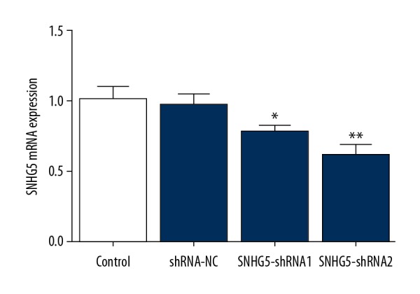
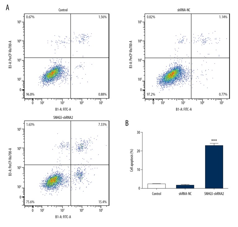
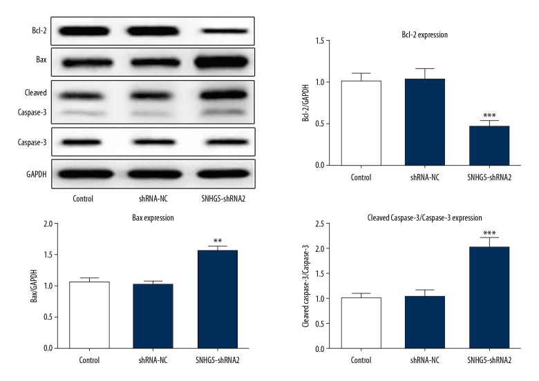
BACKGROUND Human chronic myelogenous leukemia (CML) is a hematopoietic stem cell disorder with high malignant and invasive activity. lncRNA SNHG5 has been reported to be upregulated in CML. However, whether it affects the proliferation, differentiation, and apoptosis in CML cells is still unknown. This study investigated the role of SNHG5 in CML and revealed the potential mechanism. MATERIAL AND METHODS K562 cells were transfected with shRNA, and the expression level of SNHG5 was assessd by quantitative RT-PCR. The proliferation ability was determined by CCK-8 assay. Western blot analysis was performed to detect protein expressions related to cell proliferation, differentiation, and apoptosis. Cell apoptosis rate was analyzed by flow cytometry. The DNA methylation level was determined by methylation-specific PCR (MSP). RESULTS Our results show that inhibition of SNHG5 induced by RNA interference significantly inhibits K562 cells proliferation and induces cell differentiation with the increased expression of CD42b, CD11b, CD14, GATA-1, and ß-globin. Flow cytometry analysis indicated that inhibition of SNHG5 significantly induced cell apoptosis with decreased expression of Bcl-2 and increased expression of Bax and cleaved capase-3. Additionally, Western blot and MSP analyses confirmed that inhibition of SNHG5 increased the expression of DR4 gene through suppressing its methylation. CONCLUSIONS Inhibition of SNHG5 suppressed K562 cell proliferation through inducing the differentiation and apoptosis by inhibiting methylation of DR4. Therefore, downregulated SNHG5 could play a key role in CML progression, and might provide a new strategy for the treatment of CML.
Conflict of interest statement
None.
Figures


Similar articles
-
Latest Advances of Long Non-Coding RNA SNHG5 in Human Cancers.Onco Targets Ther. 2020 Jul 1;13:6393-6403. doi: 10.2147/OTT.S252750. eCollection 2020. Onco Targets Ther. 2020. PMID: 32753882 Free PMC article. Review.
-
LncRNA HAND2-AS1 inhibits proliferation and promotes apoptosis of chronic myeloid leukemia cells by sponging with micRNA-1275.Eur Rev Med Pharmacol Sci. 2019 Mar;23(5):2103-2111. doi: 10.26355/eurrev_201903_17254. Eur Rev Med Pharmacol Sci. 2019. PMID: 30915755
-
Silencing of suppressor of cytokine signaling-3 due to methylation results in phosphorylation of STAT3 in imatinib resistant BCR-ABL positive chronic myeloid leukemia cells.Asian Pac J Cancer Prev. 2014;15(11):4555-61. doi: 10.7314/apjcp.2014.15.11.4555. Asian Pac J Cancer Prev. 2014. PMID: 24969884
-
Long noncoding RNA MEG3 inhibits proliferation of chronic myeloid leukemia cells by sponging microRNA21.Biomed Pharmacother. 2018 Aug;104:181-192. doi: 10.1016/j.biopha.2018.05.047. Epub 2018 May 15. Biomed Pharmacother. 2018. PMID: 29772439
-
LncRNA SNHG5: A new budding star in human cancers.Gene. 2020 Jul 30;749:144724. doi: 10.1016/j.gene.2020.144724. Epub 2020 May 1. Gene. 2020. PMID: 32360843 Review.
Cited by
-
Aberrant LncRNA Expression in Leukemia.J Cancer. 2020 Apr 27;11(14):4284-4296. doi: 10.7150/jca.42093. eCollection 2020. J Cancer. 2020. PMID: 32368311 Free PMC article. Review.
-
Molecular-Genetic Biomarkers of Diabetic Macular Edema.J Clin Med. 2024 Dec 5;13(23):7426. doi: 10.3390/jcm13237426. J Clin Med. 2024. PMID: 39685883 Free PMC article. Review.
-
Novel Insights on Ferroptosis Modulation as Potential Strategy for Cancer Treatment: When Nature Kills.Antioxid Redox Signal. 2024 Jan;40(1-3):40-85. doi: 10.1089/ars.2022.0179. Epub 2023 Jun 28. Antioxid Redox Signal. 2024. PMID: 37132605 Free PMC article. Review.
-
Upregulation of lncRNA147410.3 in the Brain of Mice With Chronic Toxoplasma Infection Promoted Microglia Apoptosis by Regulating Hoxb3.Front Cell Neurosci. 2021 May 18;15:648047. doi: 10.3389/fncel.2021.648047. eCollection 2021. Front Cell Neurosci. 2021. PMID: 34084127 Free PMC article.
-
Latest Advances of Long Non-Coding RNA SNHG5 in Human Cancers.Onco Targets Ther. 2020 Jul 1;13:6393-6403. doi: 10.2147/OTT.S252750. eCollection 2020. Onco Targets Ther. 2020. PMID: 32753882 Free PMC article. Review.
References
-
- Kaleem B, Shahab S, Ahmed N, Shamsi TS. Chronic myeloid leukemia – prognostic value of mutations. Asian Pac J Cancer Prev. 2015;16(17):7415–23. - PubMed
-
- Usmani SZ, Yunus SA, Jamal Y. Overview of chronic myeloid leukemia patients in Pakistan in the pre-imatanib era. Asian Pac J Cancer Prev. 2009;10(6):1039–40. - PubMed
MeSH terms
Substances
LinkOut - more resources
Full Text Sources
Medical
Research Materials
