. 2019 Sep 10;10(9):662.
doi: 10.1038/s41419-019-1919-0.

ATF6 regulates the development of chronic pancreatitis by inducing p53-mediated apoptosis

Affiliations

ATF6 regulates the development of chronic pancreatitis by inducing p53-mediated apoptosis

Lei Zhou et al. Cell Death Dis. .

Abstract

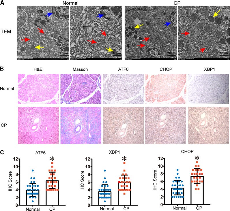
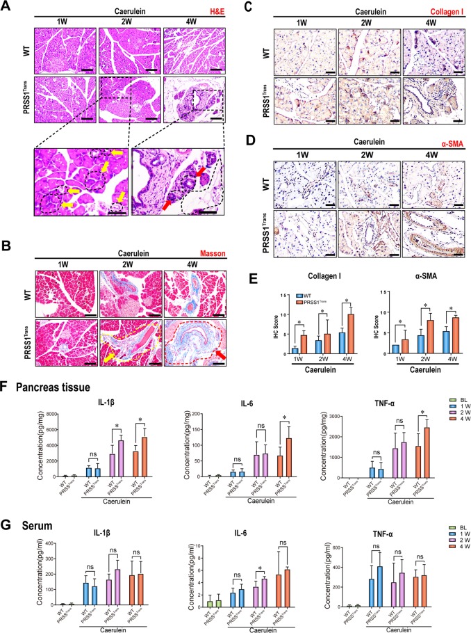
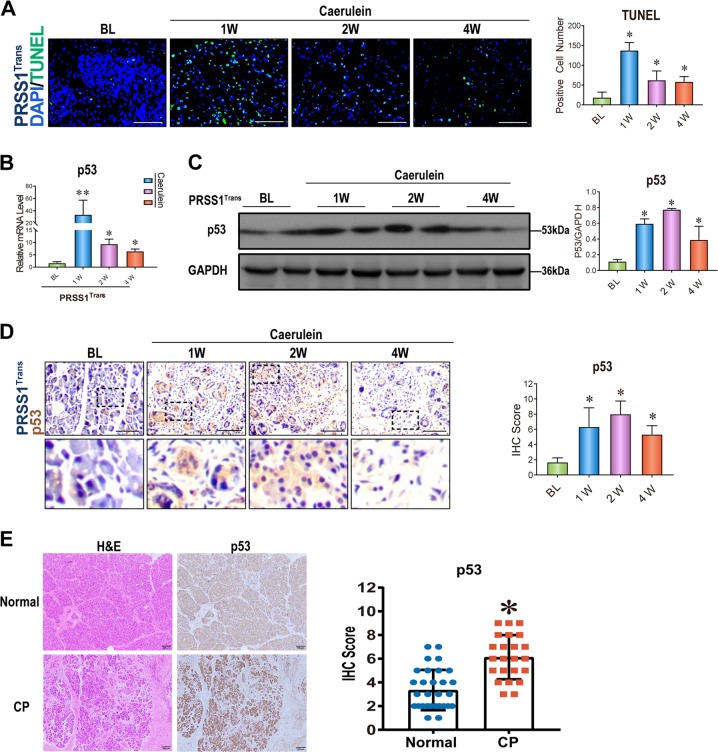
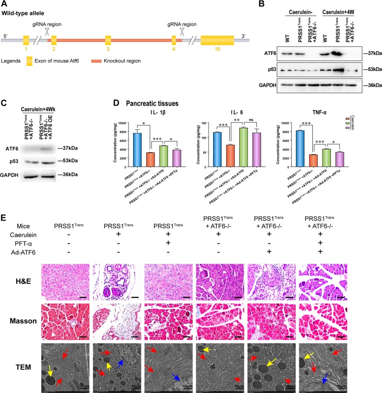
Chronic pancreatitis (CP) is a progressive, recurrent inflammatory disorder of the pancreas. Initiation and progression of CP can result from serine protease 1 (PRSS1) overaccumulation and the ensuing endoplasmic reticulum (ER) stress. However, how ER stress pathways regulate the development and progression of CP remains poorly understood. In the present study we aimed to elucidate the ER stress pathway involved in CP. We found high expression of the ER stress marker genes ATF6, XBP1, and CHOP in human clinical specimens. A humanized PRSS1 transgenic mouse was established and treated with caerulein to mimic the development of CP, as evidenced by pathogenic alterations, collagen deposition, and increased expression of the inflammatory factors IL-6, IL-1β, and TNF-α. ATF6, XBP1, and CHOP expression levels were also increased during CP development in this model. Acinar cell apoptosis was also significantly increased, accompanied by upregulated p53 expression. Inhibition of ATF6 or p53 suppressed the expression of inflammatory factors and progression of CP in the mouse model. Finally, we showed that p53 expression could be regulated by the ATF6/XBP1/CHOP axis to promote the development of CP. We therefore conclude that ATF6 signalling regulates CP progression by modulating pancreatic acinar cell apoptosis, which provides a target for ER stress-based diagnosis and treatment of CP.


Conflict of interest statement

The authors declare that they have no conflict of interest.

Figures

Fig. 1
Fig. 1. ER destruction in human CP tissues accompanied by high expression of the ER stress-responsive factors ATF6/XBP1/CHOP.
a The morphology of human normal pancreatic tissue and chronic pancreatitis (CP) tissue was observed by transmission electron microscopy (TEM). Red arrows (↑): endoplasmic reticulum; yellow arrows (↑): zymogen granule; blue arrows (↑): cell nucleus. b Haematoxylin and eosin (H&E) and Masson’s trichrome staining of pancreatic tissues and ATF6, CHOP, and XBP1 expression determined by immunohistochemistry (IHC) in CP patients and healthy volunteers. c The IHC score was used to quantify the relative expression of ATF6, CHOP, and XBP1. ATF6 activating transcription factor 6, CHOP C/EBP-homologous protein, ER endoplasmic reticulum, XBP1 X-box-binding protein 1. *P < 0.05
Fig. 2
Fig. 2. CP model established by caerulein injection in PRSS1 transgenic mice (PRSSTrans).
a Histological evaluation of pancreatic tissues collected from chronic pancreatitis (CP) model mice and wild-type (WT) mice by haematoxylin and eosin (H&E) staining. b Analysis of collagen deposition by Masson’s trichrome staining (Red (↑) and yellow arrows (↑) indicate increased fibrosis in pancreatic tissues from PRSS1 transgenic mice at 2 and 4 weeks post-caerulein injection in pancreatic tissues from PRSS1 transgenic mice, respectively.) and c collagen I levels and d α-smooth muscle actin (α-SMA) levels by immunohistochemistry (IHC) in pancreatic tissues from CP model mice and WT mice sacrificed 1, 2, or 4 weeks post-caerulein injection. e IHC histological scores of pancreatic tissues in CP model mice after finishing caerulein treatment. The levels of IL-1β, IL-6, and TNF-α in pancreatic tissues (f) and serum (g) from WT and PRSS1 transgenic mice treated with caerulein for CP induction, as determined by ELISA and quantitative RT-PCR. PRSS1 serine protease 1, α-SMA α-smooth muscle actin, IL-1β interleukin 1β, IL-6 interleukin 6, TNF-α tumour necrosis factor-α, BL baseline, W weeks, ns no significant difference; *P < 0.05
Fig. 3
Fig. 3. ER stress responses in PRSS1 transgenic mice treated with caerulein.
a Morphological changes in the endoplasmic reticulum (ER) in response to caerulein treatment in PRSS1 transgenic mice were determined by transmission electron microscopy. Red arrows (↑): endoplasmic reticulum; yellow arrows (↑): zymogen granule; blue arrows (↑): cell nucleus. ATF6, XBP1, and CHOP gene (b) and protein (c) levels in pancreatic tissues, as determined by quantitative RT-PCR and western blotting, respectively. ATF6 activating transcription factor 6, XBP1 X-box-binding protein 1, CHOP C/EBP-homologous protein, GAPDH glyceraldehyde-3-phosphate dehydrogenase, ns no significant difference, WT wild-type, BL baseline, W weeks, ns no significant difference; *P < 0.05 and **P < 0.01
Fig. 4
Fig. 4. Acinar apoptosis and p53 expression in CP.
a Transferase-mediated d-UTP nick-end-labelling (TUNEL) assay in PRSS1 transgenic mice treated with caerulein for 1, 2, and 4 weeks. b p53 mRNA levels in pancreatic tissues from PRSS1 transgenic mice treated with caerulein to induce chronic pancreatitis (CP). Mice were sacrificed 1, 2, and 4 weeks after treatment, and the mRNA levels were determined by quantitative RT-PCR. p53 protein levels in pancreatic tissues from PRSS1 transgenic mice treated with caerulein by Western blotting (c) and immunohistochemistry (IHC) (d). e Haematoxylin and eosin (h, e) and p53 IHC in CP patients and healthy volunteers. BL baseline, W weeks; *P < 0.05
Fig. 5
Fig. 5. Regulation of ER stress by p53 during CP progression in PRSS1 transgenic mice.
a The expression of p53 in PRSS1 transgenic mice treated with pifithrin-α (PFT-α; a p53 inhibitor) and tauroursodeoxycholic acid (TUDCA; an ER stress inhibitor) at 1, 2, and 4 weeks after treatment with caerulein. b Transferase-mediated d-UTP nick-end-labelling (TUNEL) assay showing acinar cell apoptosis in caerulein-treated PRSS1 transgenic mice after PFT-α and TUDCA treatments. c p53 and α-smooth muscle actin (α-SMA) protein expression levels in pancreatic tissues from caerulein-treated PRSS1 transgenic mice with p53 inhibition by PFTα. d Pathological alteration of pancreatic tissues from caerulein-treated PRSS1 transgenic mice with p53 inhibition by PFTα. Tissues were stained with haematoxylin and eosin (H&E). e Expression of IL-1β, IL-6, and TNF-α in pancreatic tissues from caerulein-treated PRSS1 transgenic mice treated with PFT-α and TUDCA. IL-1β interleukin 1β, IL-6 interleukin 6, TNF-α tumour necrosis factor-α, BL baseline, W weeks, ns no significant difference; *P < 0.05 and **P < 0.01
Fig. 6
Fig. 6. Regulation of p53 expression and CP progression by ATF6 in PRSS1 acinar cells.
The expression levels of ATF6 and p53 in response to lipopolysaccharide (LPS) induction in primary acinar cells isolated from PRSS1 transgenic mice were determined by quantitative RT-PCR (a) and western blotting (b). ATF6 expression (c), inflammatory factors (d), and cell apoptosis (e) measured by quantitative RT-PCR, ELISA, and transferase-mediated d-UTP nick-end-labelling (TUNEL) assays, respectively. f Expression of ATF6 and p53 in LPS-induced ATF6-suppressed cells. shATF6 shATF6 virus, CON control group, ATF6 activating transcription factor 6, ns no significant difference; *P < 0.05 and **P < 0.01
Fig. 7
Fig. 7. Regulation of p53 expression and CP progression by ATF6 in the PRSS1 CP model.
a Schematic diagram of ATF6 knockout mice. b The expression levels of ATF6 and p53 were determined by western blotting. ATF6 and p53 protein levels (c) and IL-6, IL-1β, and TNF-α pancreatic tissues concentrations (d) in PRSS1 transgenic mice with ATF6 knockout and overexpression after induction of chronic pancreatitis (CP) for 4 weeks. e Haematoxylin and eosin (h, e) and Masson’s trichrome staining and transmission electron microscopy (TEM) images showing the pathological changes in ATF6-deficient PRSS1 transgenic mice and ATF6 deficiency in ATF6-rescued PRSS1 transgenic mice. Red arrows (↑): endoplasmic reticulum; yellow arrows (↑): zymogen granule; blue arrows (↑): cell nucleus. ATF6 activating transcription factor 6, OE overexpression, Ad-ATF6 Ad-ATF6 virus, PFT-α pifithrin-α, IL-1β interleukin 1β, IL-6 interleukin 6, TNF-α tumour necrosis factor-α. ns no significant difference, W week, *P < 0.05 and **P < 0.01

Similar articles

Cited by

References

    1. Chen JM, Férec C. Chronic pancreatitis: genetics and pathogenesis. Annu. Rev. Genomics Hum. Genet. 2009;10:63–87. doi: 10.1146/annurev-genom-082908-150009. - DOI - PubMed
    1. Chen JM, Férec C. Genetics and pathogenesis of chronic pancreatitis: The 2012 update. Clin. Res. Hepatol. Gastroenterol. 2012;36:334–340. doi: 10.1016/j.clinre.2012.05.003. - DOI - PubMed
    1. Coté GA, et al. Alcohol and smoking as risk factors in an epidemiology study of patients with chronic pancreatitis. Clin. Gastroenterol. Hepatol. 2011;9:266–273. doi: 10.1016/j.cgh.2010.10.015. - DOI - PMC - PubMed
    1. Bekkali NL, et al. Pancreatoscopy-directed electrohydraulic lithotripsy for pancreatic ductal stones in painful chronic pancreatitis using spyglass. Pancreas. 2017;46:528. doi: 10.1097/MPA.0000000000000790. - DOI - PubMed
    1. Hegyi E, Sahin-Tóth M. Genetic risk in chronic pancreatitis: the trypsin-dependent pathway. Digest. Dis. Sci. 2017;62:1692–1701. doi: 10.1007/s10620-017-4601-3. - DOI - PMC - PubMed

Publication types