. 2019 Sep 10;10(9):659.
doi: 10.1038/s41419-019-1879-4.

Exosome-mediated uptake of mast cell tryptase into the nucleus of melanoma cells: a novel axis for regulating tumor cell proliferation and gene expression

Affiliations

Exosome-mediated uptake of mast cell tryptase into the nucleus of melanoma cells: a novel axis for regulating tumor cell proliferation and gene expression

Fabio Rabelo Melo et al. Cell Death Dis. .

Abstract

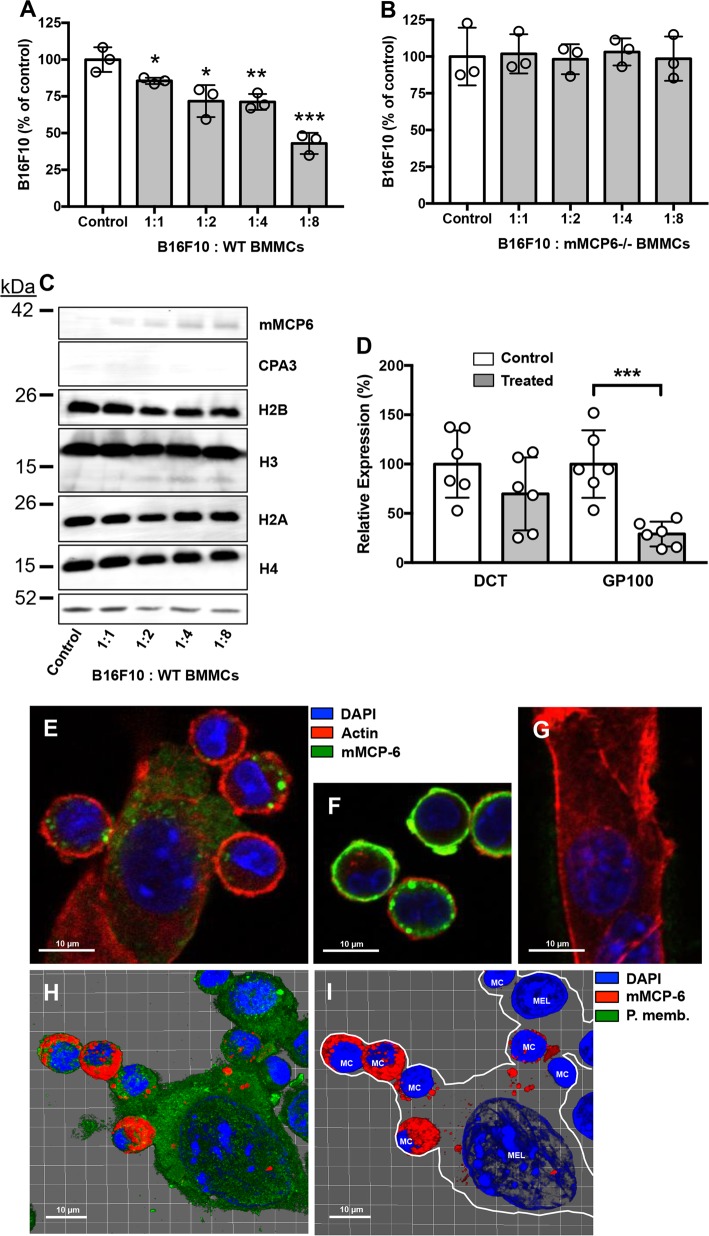
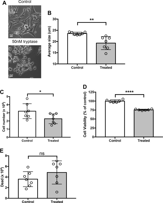
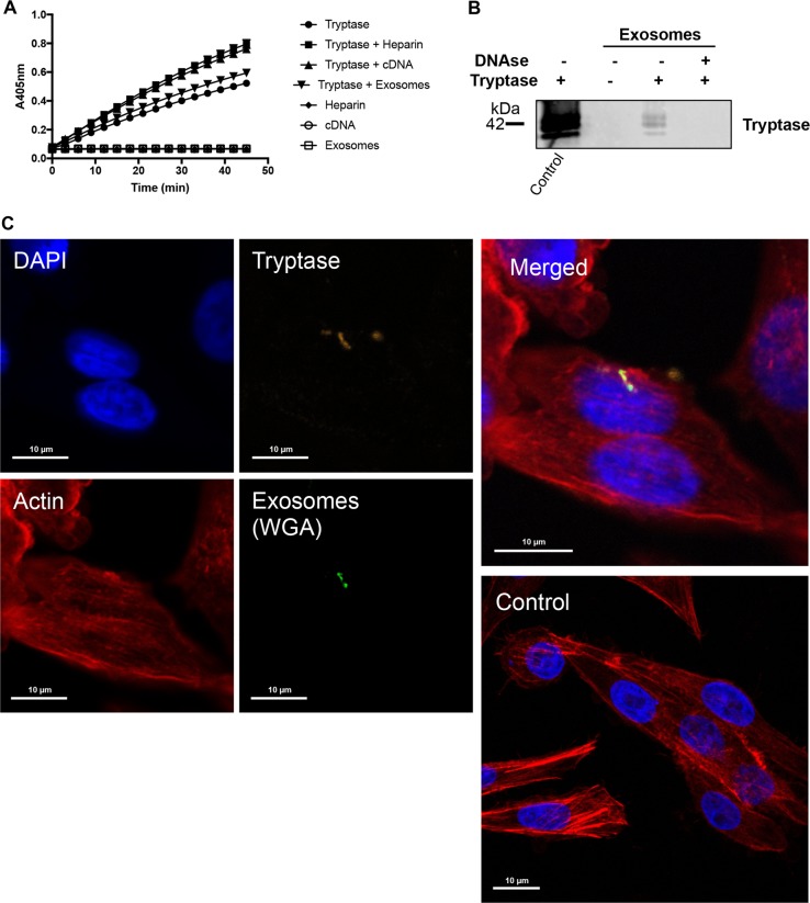
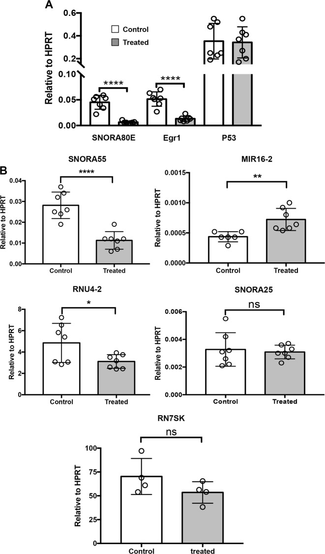
It is well established that mast cell accumulation accompanies most malignancies. However, the knowledge of how mast cells functionally impact on tumors is still rudimentary. Here we addressed this issue and show that mast cells have anti-proliferative activity on melanoma cells and that this effect is dependent on tryptase, a tetrameric protease stored in mast cell granules. Mechanistically, tryptase was found to be endocytosed by melanoma cells as cargo of DNA-coated exosomes released from melanoma cells, followed by transport to the nucleus. In the nucleus, tryptase executed clipping of histone 3 and degradation of Lamin B1, accompanied by extensive nuclear remodeling. Moreover, tryptase degraded hnRNP A2/B1, a protein involved in mRNA stabilization and interaction with non-coding RNAs. This was followed by downregulated expression of the oncogene EGR1 and of multiple non-coding RNAs, including oncogenic species. Altogether, these findings establish a new principle for regulation of tumor cell proliferation.


Conflict of interest statement

The authors declare that they have no conflict of interest.

Figures

Fig. 1
Fig. 1. Mast cell tryptase suppresses melanoma cell expansion and deliver tryptase into the nucleus of melanoma cells.
WT (a) or tryptase-deficient (mMCP6−/−) (b) mouse mast cells were co-cultured with mouse melanoma cells (B16F10) at the ratios indicated, followed by quantification of viable cells using Trypan Blue exclusion. c After co-culture, melanoma cells were separated from mast cells and were analyzed by Western blotting for cell-associated tryptase (mMCP6), CPA3, and core histones H2B, H3, H2A, and H4. Actin was used as loading control. d After co-culture of mast cells and melanoma cells, cells were harvested and analyzed by qPCR for expression of melanoma markers DCT and GP100. The displayed results are from individual experiments, representative of at least three experiments. Data in (a) and (d) are given as mean values ± SEM; *p ≤ 0.05, **p ≤ 0.01, ***p ≤ 0.001. e Confocal microscopy image showing physical contact between mast cells (MC) and melanoma cells (Mel) after co-culture. f, g Controls showing mast cells (f) and melanoma cells (g) alone. h, i Confocal images were analyzed with IMARIS software to generate 3-D-images of the interaction between mast cells and melanoma cells. Note the presence of mast cell tryptase in the nucleus of melanoma cells after mast cell:melanoma cell co-culture
Fig. 2
Fig. 2. Human recombinant tryptase affects human melanoma cell phenotype and expansion.
Human melanoma cells (MEL526) were incubated for 48 hours with 50 nM human recombinant tryptase. a Microscopy analysis revealing gross morphological alterations in melanoma cell treated with tryptase. b Assessment of cellular size showing that tryptase causes a reduction in melanoma cell size. c, d Quantification of viable cells using Trypan Blue exclusion (c) and CellTiter-Blue® viability probe (d). e Dead cells were quantified using Trypan Blue positivity. Data in (be) are given as mean values ± SEM; *p ≤ 0.05, **p ≤ 0.01, ****p ≤ 0.0001
Fig. 3
Fig. 3. Tryptase causes decreased proliferation of melanoma cells.
Melanoma cells (MEL526) were incubated for 48 hours with 50 nM human tryptase. a After incubation, melanoma cells were stained with Annexin V/Draq7 to monitor apoptosis (Annexin V+/Draq7−) and necrosis (Annexin V+/Draq7+). Note that tryptase does not induce apoptosis/necrosis in melanoma cells. b Quantification of the Annexin V/Draq7 staining. c Tryptase-treated melanoma cells (MEL526) were assessed for proliferation through EdU staining. Note the decreased EdU staining after treatment of melanoma cells with tryptase. dg Quantification of EdU staining of MEL526 (d), MM466 (e), MM253 (f), and A375 (g) melanoma cells. Data in (b, dg) are given as mean values ± SEM; **p ≤ 0.01, ***p ≤ 0.001; ns, not significant
Fig. 4
Fig. 4. Tryptase is taken up into the nucleus of human melanoma cells, and affects nuclear morphology and histone processing.
a Human melanoma cells (MEL526) were incubated with human tryptase at the indicated concentrations, followed by washing of the cells and Western blot analysis for tryptase. Histone 1 (H1) was used as loading control. b Western blot analysis for core histones H2A, H2B, H3, and H4 after incubation of human melanoma cells with tryptase at the indicated concentrations for 48 h. Note the clipping of H3 after treatment of melanoma cells with tryptase. c Confocal analysis showing uptake of tryptase into melanoma cells and blocked uptake in the presence of an endocytosis inhibitor (Dynasore). d 3-D imaging using the IMARIS software showing the presence of tryptase in both the cytoplasm and nucleus of human melanoma cells (see also Supplementary Fig. 2). e DAPI staining of control and tryptase-treated human melanoma cells using super-resolution microscopy analysis. Note the extensive morphological alterations of the nucleus after tryptase treatment. Note also the presence of pores in the nuclear envelope after treatment with tryptase
Fig. 5
Fig. 5. Tryptase is taken up into human melanoma cells as cargo of melanoma cell-derived exosomes.
a Human tryptase was incubated with heparin, cDNA or purified melanoma-derived exosomes as indicated, followed by assessment of enzymatic activity using a chromogenic substrate (S-2288). Note that the symbols representing samples without tryptase (Heparin, cDNA, exosomes) are superimposed and show no detectable activity against S-2288. b Purified melanoma cell-derived exosomes (~4 × 107 exosomes) were either non-treated or digested with DNAse and then incubated with or without tryptase (as indicated), followed by washing and detection of bound tryptase by Western blot analysis. Purified tryptase was included as positive control (left lane). c Purified melanoma cell-derived exosomes (~2.5 × 107 exosomes) were labeled with WGA, incubated with purified tryptase, washed and then incubated with human melanoma cells for 6 h, followed by confocal microscopy analysis. Note the presence of tryptase:WGA double-positive exosomes in the nucleus and cytoplasm of the melanoma cells
Fig. 6
Fig. 6. Tryptase degrades nuclear hnRNP A2/B1 and Lamin B1 in human melanoma cells.
a Nuclear extracts were prepared from human melanoma cells and were incubated with tryptase as indicated, followed by SDS-PAGE analysis and Coomassie staining. The major bands affected by tryptase (arrows) were identified as hnRNP A2/B1 and Lamin B1, respectively. bd Western blot analysis of hnRNP A2/B1 and Lamin B1 in tryptase-treated nuclear extracts. Quantification of the Lamin B1 and hnRNP A2/B1 bands by densitometric scanning is shown in (c) and (d), respectively. eg Western blot analysis of hnRNP A2/B1 and Lamin B1 in tryptase-treated cells. Quantification of the Lamin B1 and hnRNP A2/B1 bands by densitometric scanning is shown in (f) and (g), respectively. h Recombinant hnRNP A2/B1 was treated with tryptase, followed by SDS-PAGE and Coomassie staining
Fig. 7
Fig. 7. Tryptase affects the expression of non-coding RNAs and EGR1 in human melanoma cells.
Human melanoma cells (MEL526) were incubated with 50 nM tryptase for 48 h, followed by qPCR analysis of the expression of SNORA80E, EGR1, P53, SNORA55, MIR16–2, RNU4–2, SNORA25, and RN7SK as indicated. Data are given as mean values ± SEM; *p ≤ 0.05, **p ≤ 0.01, ****p ≤ 0.0001

Similar articles

Cited by

References

    1. Metcalfe DD, Baram D, Mekori YA. Mast cells. Physiol. Rev. 1997;77:1033–1079. doi: 10.1152/physrev.1997.77.4.1033. - DOI - PubMed
    1. Gurish MF, Austen KF. Developmental origin and functional specialization of mast cell subsets. Immunity. 2012;37:25–33. doi: 10.1016/j.immuni.2012.07.003. - DOI - PubMed
    1. Galli SJ, Nakae S, Tsai M. Mast cells in the development of adaptive immune responses. Nat. Immunol. 2005;6:135–142. doi: 10.1038/ni1158. - DOI - PubMed
    1. Ribatti D, Crivellato E. The controversial role of mast cells in tumor growth. Int. Rev. Cell Mol. Biol. 2009;275:89–131. doi: 10.1016/S1937-6448(09)75004-X. - DOI - PubMed
    1. Dalton DK, Noelle RJ. The roles of mast cells in anticancer immunity. Cancer Immunol. Immunother. 2012;61:1511–1520. doi: 10.1007/s00262-012-1246-0. - DOI - PMC - PubMed

Publication types