. 2019 Sep 10;9(1):12957.
doi: 10.1038/s41598-019-49260-4.

Genomic and developmental characterisation of a novel bunyavirus infecting the crustacean Carcinus maenas

Affiliations

Genomic and developmental characterisation of a novel bunyavirus infecting the crustacean Carcinus maenas

Jamie Bojko et al. Sci Rep. .

Abstract

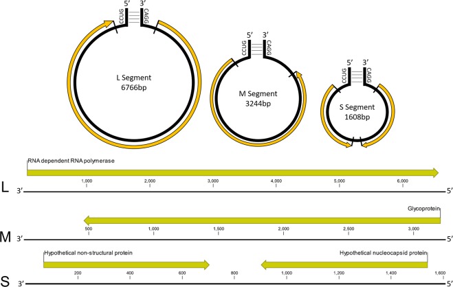
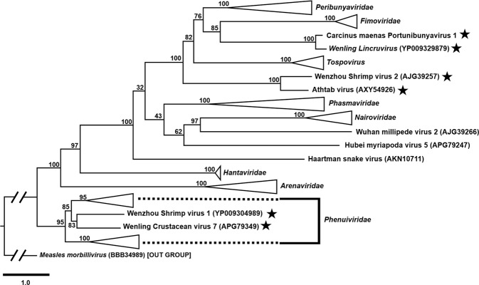
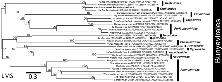
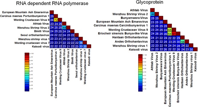
Carcinus maenas is in the top 100 globally invasive species and harbours a wide diversity of pathogens, including viruses. We provide a detailed description for a novel bunyavirus (Carcinus maenas Portunibunyavirus 1) infecting C. maenas from its native range in the Faroe Islands. The virus genome is tripartite, including large (L) (6766 bp), medium (M) (3244 bp) and small (S) (1608 bp) negative sense, single-stranded RNA segments. Individual genomic segments are flanked by 4 bp regions of similarity (CCUG). The segments encode an RNA-dependent RNA-polymerase, glycoprotein, non-structural protein with a Zinc-Finger domain and a nucleoprotein. Most show highest identity to the 'Wenling Crustacean Virus 9' from an unidentified crustacean host. Phylogenomics of crustacean-infecting bunyaviruses place them across multiple bunyavirus families. We discuss the diversity of crustacean bunyaviruses and provide an overview of how these viruses may affect the health and survival of crustacean hosts, including those inhabiting niches outside of their native range.


Conflict of interest statement

The authors declare no competing interests.

Figures

Figure 1
Figure 1
Electron micrographs of the developmental cycle for ‘Carcinus maenas Portunibunyavirus 1’. (A) Single membrane viruses exit/enter the cell via cytosis of the cell membrane (black arrows) and enter the cell with a double membrane (white arrow) vesicles (V) including developing viral particles are present in section. (B) A lower magnification image shows an infected cell, among adjacent cells, with multiple vesicles containing developing virions. The nucleus (N) of an adjacent cell can be identified. (C) An image of an early developing vacuole with electron dense material, predicted to be viral RNA undergoing translation/transcription. (D) Virions are assembled. Vacuoles with late stage virions (V1) and pre-full-assembly virions are present in section (V2). (E) In some infected cells a paracrystalline array was noted to form in the cytoplasm of the host. F) As virions leave the cell, they shed their second membrane (black arrow). (G) Finally, infective virions enter the interstitial space between cells and aggregate before moving on to adjacent cells.
Figure 2
Figure 2
A graphical representation of the putative viral development for ‘Carcinus maenas Portunibunyavirus 1’ based on transmission electron micrographs attained from infected gill tissue. Infective stages enter the cell through endocytosis (1/Fig. 1A), providing them with a second membrane. This membrane is then predicted to shed (2), allowing release of viral -ssRNA. The genetic material then interacts with the cell, possibly through multiple pathways yet to be defined (3) but result in a series of large vacuoles where viral assembly occurs (4/Fig. 1C). After assembly, viral particles move from the assembly vesicle (5/Fig. 1D,G) and exit the cell (6/Fig. 1A). In addition to the classical bunyaviral development cycle, multiple occurrences of viroplasm development occur, suggesting a build-up of virions in the cytoplasm of the host cell which may rupture to result in virion release (“VIROPLASM”) (Fig. 1E). The diagram is not to cellular scale.
Figure 3
Figure 3
The annotated -ssRNA genome of ‘Carcinus maenas Portunibunyavirus 1’ and graphical representation of the three -ssRNA genomic segments with annotation for the bunyavirus-associated open reading frames. The predicted protein is listed above the annotation and the complementary bunyaviral ends are represented.
Figure 4
Figure 4
A phylogenetic tree developed from the predicted RdRp proteins of 97 bunyaviruses and an outgroup (Mononegavirales: Measles morbillivirus). The protein sequence data were aligned in Geneious using MAFFT default protocol. The tree was developed using IQ-tree. The outgroup or corresponding viral family are highlighted on the tree and the crustacean-infecting bunyaviruses are identified with a star. The FASTA file used to create the tree is available in the supplementary information (Suppl. File 1).
Figure 5
Figure 5
A concatenated phylogenetic tree developed from the L, M and S segment (LMS) proteins of 29 bunyaviruses. The protein sequence data were aligned separately in Geneious using MAFFT default protocol and then merged. The tree was developed from the merged data using IQ-tree. The corresponding viral family within the Bunyavirales is highlighted on the tree, including the Cruliviridae and ‘Carcinus maenas Portunibunyavirus 1’ in bold. The accession numbers for the L, M and S proteins, according to the NCBI repository, are presented after each viral isolate used in the tree.
Figure 6
Figure 6
Protein distance matrix for the crustacean-infecting bunyaviruses and representatives from the closest relatable Bunyavirales families.
Figure 7
Figure 7
RNA-dependent-RNA-polymerase conserved domain conservation and amino acid sequence similarity using comparative methods explored by Amroun et al.. Conservation of all amino acids (or amino acids with similar properties) across the sequences are highlighted in black. Partial amino acid conservation across most sequences is represented in grey. Carcinus maenas Portunibunyavirus 1 (CmPBV1), Wenling Crustacean Virus 9 (WCV9), Athtab virus (AV), Wenling Crustacean Virus 7 (WCV7), Wenzhou Shrimp Virus 1 (WSV1), Wenzhou Shrimp Virus 2 (WSV2), and Hantaan virus (Hantaan) are each compared. Data and images acquired from Geneious MAFFT alignment.

Similar articles

Cited by

  • A Novel Bunyavirus Discovered in Oriental Shrimp (Penaeus chinensis).
    Dong X, Hu T, Ren Y, Meng F, Li C, Zhang Q, Chen J, Song J, Wang R, Shi M, Li J, Zhao P, Li C, Tang KFJ, Cowley JA, Shi W, Huang J. Dong X, et al. Front Microbiol. 2021 Nov 24;12:751112. doi: 10.3389/fmicb.2021.751112. eCollection 2021. Front Microbiol. 2021. PMID: 34899637 Free PMC article.
  • Family Level Phylogenies Reveal Relationships of Plant Viruses within the Order Bunyavirales.
    Herath V, Romay G, Urrutia CD, Verchot J. Herath V, et al. Viruses. 2020 Sep 10;12(9):1010. doi: 10.3390/v12091010. Viruses. 2020. PMID: 32927652 Free PMC article.
  • Mycosis is a Disease State Encountered Rarely in Shore Crabs, Carcinus maenas.
    Davies CE, Malkin SH, Thomas JE, Batista FM, Rowley AF, Coates CJ. Davies CE, et al. Pathogens. 2020 Jun 11;9(6):462. doi: 10.3390/pathogens9060462. Pathogens. 2020. PMID: 32545349 Free PMC article.
  • 2021 Taxonomic update of phylum Negarnaviricota (Riboviria: Orthornavirae), including the large orders Bunyavirales and Mononegavirales.
    Kuhn JH, Adkins S, Agwanda BR, Al Kubrusli R, Alkhovsky SV, Amarasinghe GK, Avšič-Županc T, Ayllón MA, Bahl J, Balkema-Buschmann A, Ballinger MJ, Basler CF, Bavari S, Beer M, Bejerman N, Bennett AJ, Bente DA, Bergeron É, Bird BH, Blair CD, Blasdell KR, Blystad DR, Bojko J, Borth WB, Bradfute S, Breyta R, Briese T, Brown PA, Brown JK, Buchholz UJ, Buchmeier MJ, Bukreyev A, Burt F, Büttner C, Calisher CH, Cao M, Casas I, Chandran K, Charrel RN, Cheng Q, Chiaki Y, Chiapello M, Choi IR, Ciuffo M, Clegg JCS, Crozier I, Dal Bó E, de la Torre JC, de Lamballerie X, de Swart RL, Debat H, Dheilly NM, Di Cicco E, Di Paola N, Di Serio F, Dietzgen RG, Digiaro M, Dolnik O, Drebot MA, Drexler JF, Dundon WG, Duprex WP, Dürrwald R, Dye JM, Easton AJ, Ebihara H, Elbeaino T, Ergünay K, Ferguson HW, Fooks AR, Forgia M, Formenty PBH, Fránová J, Freitas-Astúa J, Fu J, Fürl S, Gago-Zachert S, Gāo GF, García ML, García-Sastre A, Garrison AR, Gaskin T, Gonzalez JJ, Griffiths A, Goldberg TL, Groschup MH, Günther S, Hall RA, Hammond J, Han T, Hepojoki J, Hewson R, Hong J, Hong N, Hongo S, Horie M, Hu JS, Hu T, Hughes HR, Hüttner F, Hyndman TH, Ilyas M, Jalkanen R, Jiāng D, Jonson GB, Junglen S, Kadono F, Ka… See abstract for full author list ➔ Kuhn JH, et al. Arch Virol. 2021 Dec;166(12):3513-3566. doi: 10.1007/s00705-021-05143-6. Arch Virol. 2021. PMID: 34463877 Free PMC article.
  • Enormous diversity of RNA viruses in economic crustaceans.
    Dong X, Meng F, Zhou C, Li J, Hu T, Wang Y, Wang G, Luo J, Li X, Liu S, Huang J, Shi W. Dong X, et al. mSystems. 2024 Oct 22;9(10):e0101624. doi: 10.1128/msystems.01016-24. Epub 2024 Sep 27. mSystems. 2024. PMID: 39329483 Free PMC article.

References

    1. Guterres A, de Oliveira RC, Fernandes J, de Lemos ERS, Schrago CG. New bunya-like viruses: Highlighting their relations. Infect. Genet. Evol. 2017;49:164–173. doi: 10.1016/j.meegid.2017.01.019. - DOI - PubMed
    1. Maes P, et al. Taxonomy of the family Arenaviridae and the order Bunyavirales: update 2018. Arch. Virol. 2018;163:2295–2310. doi: 10.1007/s00705-018-3843-5. - DOI - PubMed
    1. Maes P, et al. Taxonomy of the order Bunyavirales: second update 2018. Arch Virol. 2019;164:927–941. doi: 10.1007/s00705-018-04127-3. - DOI - PMC - PubMed
    1. Cui N, et al. Severe fever with thrombocytopenia syndrome bunyavirus-related human encephalitis. J. Infect. 2015;70:52–59. doi: 10.1016/j.jinf.2014.08.001. - DOI - PubMed
    1. Ling J, et al. Genetic analyses of Seoul hantavirus genome recovered from rats (Rattus norvegicus) in the Netherlands unveils diverse routes of spread into Europe. J. Med. Virol. 2019;91:724–730. doi: 10.1002/jmv.25390. - DOI - PubMed

Publication types