

Pro-Cellular Exhaustion Markers are Associated with Splenic Microarchitecture Disorganization and Parasite Load in Dogs with Visceral Leishmaniasis
- PMID: 31506501
- PMCID: PMC6736856
- DOI: 10.1038/s41598-019-49344-1
Pro-Cellular Exhaustion Markers are Associated with Splenic Microarchitecture Disorganization and Parasite Load in Dogs with Visceral Leishmaniasis
Abstract
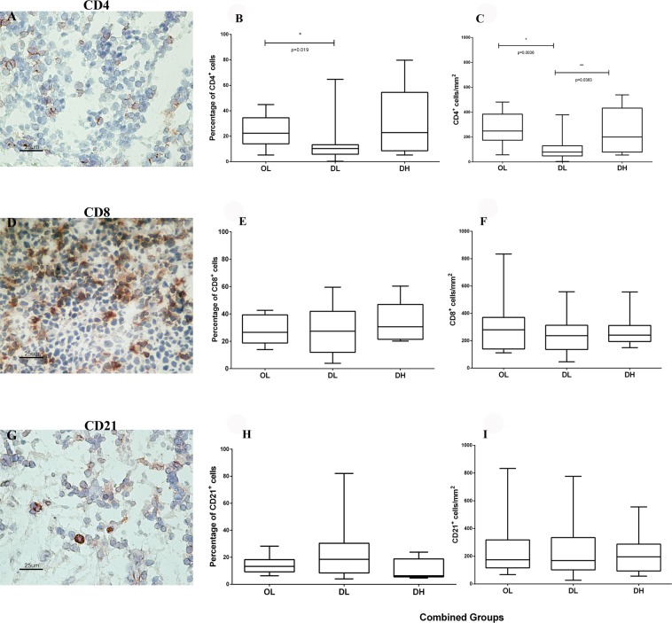
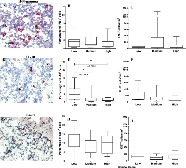
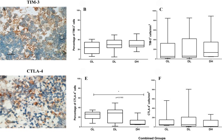
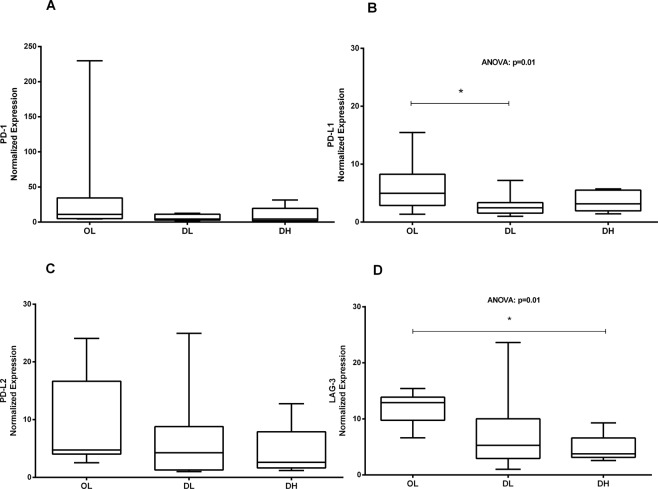
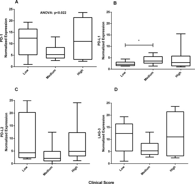
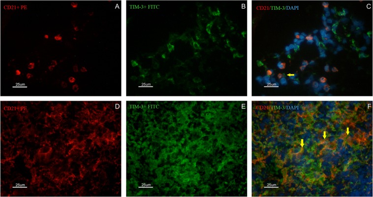
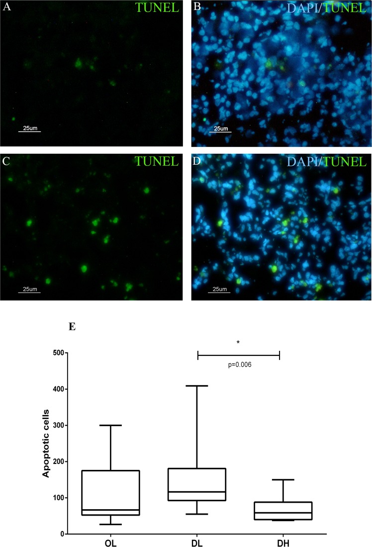
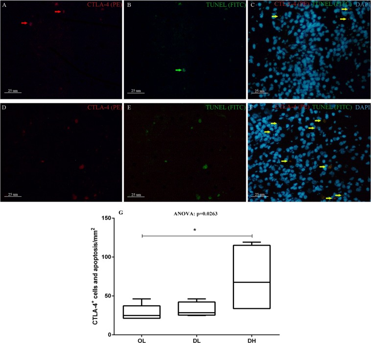
In canine visceral leishmaniasis (CVL), splenic white pulp (SWP) disorganization has been associated with disease progression, reduced cytokine and chemokine expression and failure to control the parasite load. This profile is compatible with the cellular exhaustion previously shown in human visceral leishmaniasis. The present study aimed to evaluate the in situ expression of cellular exhaustion markers and their relation to clinical signs, SWP disorganization and parasite load. Forty dogs naturally infected by Leishmania infantum were grouped according to levels of SWP organization and parasite load. SWP disorganization was associated with reductions in the periarteriolar lymphatic sheath and lymphoid follicles/mm2 and worsening of the disease. Apoptotic cells expressing CTLA-4+ increased in dogs with disorganized SWP and a high parasite load. In the same group, PD-L1 and LAG-3 gene expression were reduced. A higher number of CD21+TIM-3+ B cells was detected in disorganized spleens than in organized spleens. Apoptosis is involved in periarteriolar lymphatic sheath reduction and lymphoid follicle atrophy and is associated with CTLA-4+ cell reductions in the splenic tissue of dogs with visceral leishmaniasis (VL). Failure to control the parasite load was observed, suggesting that cell exhaustion followed by T and B cell apoptosis plays a role in the immunosuppression observed in CVL.
Conflict of interest statement
The authors declare no competing interests.
Figures
Similar articles
-
Splenic macrophage functional profile and its role in the immunopathogenesis of canine visceral leishmaniasis.Front Immunol. 2025 Jun 20;16:1617751. doi: 10.3389/fimmu.2025.1617751. eCollection 2025. Front Immunol. 2025. PMID: 40621456 Free PMC article.
-
Morphophysiological changes in the splenic extracellular matrix of Leishmania infantum-naturally infected dogs is associated with alterations in lymphoid niches and the CD4+ T cell frequency in spleens.PLoS Negl Trop Dis. 2018 Apr 20;12(4):e0006445. doi: 10.1371/journal.pntd.0006445. eCollection 2018 Apr. PLoS Negl Trop Dis. 2018. Retraction in: PLoS Negl Trop Dis. 2022 Feb 16;16(2):e0010225. doi: 10.1371/journal.pntd.0010225. PMID: 29677186 Free PMC article. Retracted.
-
Low CXCL13 expression, splenic lymphoid tissue atrophy and germinal center disruption in severe canine visceral leishmaniasis.PLoS One. 2012;7(1):e29103. doi: 10.1371/journal.pone.0029103. Epub 2012 Jan 5. PLoS One. 2012. PMID: 22242159 Free PMC article.
-
Systemic and compartmentalized immune response in canine visceral leishmaniasis.Vet Immunol Immunopathol. 2009 Mar 15;128(1-3):87-95. doi: 10.1016/j.vetimm.2008.10.307. Epub 2008 Oct 17. Vet Immunol Immunopathol. 2009. PMID: 19054576 Review.
-
Canine visceral leishmaniasis biomarkers and their employment in vaccines.Vet Parasitol. 2019 Jul;271:87-97. doi: 10.1016/j.vetpar.2019.05.006. Epub 2019 May 20. Vet Parasitol. 2019. PMID: 31303211 Review.
Cited by
-
Phenotypical Characterization of Spleen Remodeling in Murine Experimental Visceral Leishmaniasis.Front Immunol. 2020 Apr 15;11:653. doi: 10.3389/fimmu.2020.00653. eCollection 2020. Front Immunol. 2020. PMID: 32351510 Free PMC article.
-
Splenic macrophage functional profile and its role in the immunopathogenesis of canine visceral leishmaniasis.Front Immunol. 2025 Jun 20;16:1617751. doi: 10.3389/fimmu.2025.1617751. eCollection 2025. Front Immunol. 2025. PMID: 40621456 Free PMC article.
-
Splenic Transcriptional Responses in Severe Visceral Leishmaniasis: Impaired Leukocyte Chemotaxis and Cell Cycle Arrest.Front Immunol. 2021 Nov 5;12:716314. doi: 10.3389/fimmu.2021.716314. eCollection 2021. Front Immunol. 2021. PMID: 34804009 Free PMC article.
-
Cytokines and splenic remodelling during Leishmania donovani infection.Cytokine X. 2020 Sep 1;2(4):100036. doi: 10.1016/j.cytox.2020.100036. eCollection 2020 Dec. Cytokine X. 2020. PMID: 33604560 Free PMC article.
-
Leishmania Parasites Drive PD-L1 Expression in Mice and Human Neutrophils With Suppressor Capacity.Front Immunol. 2021 Jun 15;12:598943. doi: 10.3389/fimmu.2021.598943. eCollection 2021. Front Immunol. 2021. PMID: 34211455 Free PMC article.
References
Publication types
MeSH terms
Substances
LinkOut - more resources
Full Text Sources
Molecular Biology Databases
Research Materials
