. 2019 Sep 10;9(1):13008.
doi: 10.1038/s41598-019-49477-3.

Germline variability and tumor expression level of ribosomal protein gene RPL28 are associated with survival of metastatic colorectal cancer patients

Affiliations

Germline variability and tumor expression level of ribosomal protein gene RPL28 are associated with survival of metastatic colorectal cancer patients

Adrien Labriet et al. Sci Rep. .

Abstract

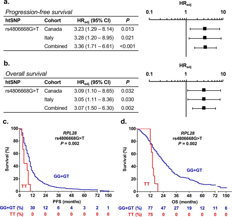
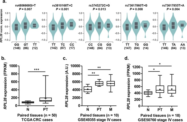
This study investigated the potential of single nucleotide polymorphisms as predictors of survival in two cohorts comprising 417 metastatic colorectal cancer (mCRC) patients treated with the FOLFIRI (folinic acid, 5-fluorouracil and irinotecan) regimen. The rs4806668G > T of the ribosomal protein gene RPL28 was associated with shorter progression-free survival and overall survival by 5 and 9 months (P = 0.002), with hazard ratios of 3.36 (P < 0.001) and 3.07 (P = 0.002), respectively. The rs4806668T allele was associated with an increased RPL28 expression in transverse normal colon tissues (n = 246, P = 0.007). RPL28 expression was higher in colorectal tumors compared to paired normal tissues by up to 124% (P < 0.001) in three independent datasets. Metastatic cases with highest RPL28 tumor expression had a reduced survival in two datasets (n = 88, P = 0.009 and n = 56, P = 0.009). High RPL28 was further associated with changes in immunoglobulin and extracellular matrix pathways. Repression of RPL28 reduced proliferation by 1.4-fold to 5.6-fold (P < 0.05) in colon cancer HCT116 and HT-29 cells. Our findings suggest that the ribosomal RPL28 protein may influence mCRC outcome.


Conflict of interest statement

The authors declare no competing interests.

Figures

Figure 1
Figure 1
RPL28 rs4806668G > T polymorphism is associated with survival  of mCRC patients treated with FOLFIRI. (a,b) Cox proportional hazards models adjusted for age and co-treatment (Canadian cohort, n = 167) and for age (Italian cohort, n = 250) showed association of rs4806668G > T with progression-free survival (PFS) and overall survival (OS) using a recessive genetic model. Tumor site did not have a statistically significant association with mCRC outcome. (c,d) Univariate Kaplan-Meier survival curves for PFS and OS according to rs4806668G > T genotype in combined cohorts. The percentage survival according to genotypes is shown under the graphs. HRadj, adjusted hazard ratio; htSNPs, haplotype-tagging single nucleotide polymorphisms; CI, confidence interval.
Figure 2
Figure 2
Rs4806668 is located in the promoter region of the RPL28 locus and is linked to several other polymorphisms (SNPs). (a) Localization of the RPL28 rs4806668G > T variant and its associated SNPs in strong linkage disequilibrium (r² > 0.80 in the European population). (b) Position of the rs4806668G > T marker and its linked SNPs relative to the translation start site of RPL28. Scores are from RegulomeDB and represent the probability for a SNP to be functional. N/A, not available. (c) Frequencies of RPL28 rs4806668G > T among different ethnic groups (Ensembl GRCh38 release 91).
Figure 3
Figure 3
Relationship between RPL28 variants, tissue type and gene expression. (a) RPL28 rs4806668G > T and linked SNPs are associated with an increased gene expression in transverse colon tissues of healthy individuals from the GTEx cohort. (b) RPL28 expression is higher in primary tumor relative to paired normal colorectal tissues (n = 50 pairs) from the TCGA cohort. (c) RPL28 expression is higher in primary colorectal tumors and liver metastases relative to paired normal colorectal tissues (n = 10 pairs) from the GSE49355 dataset. (d) RPL28 expression is higher in primary colorectal tumors and liver metastases relative to paired normal colorectal tissues (n = 18 pairs) from the GSE50760 dataset. A.U., arbitrary units; FPKM, fragments per kilobase million; N, normal tissue; PT, primary tumor tissue; M, liver metastases. *P < 0.05; **P < 0.01; ***P < 0.001.
Figure 4
Figure 4
RPL28 expression level in colorectal tumor tissues is associated with survival and changes in tumor transcriptome. (a) Kaplan-Meier curves for high and low RPL28 expression groups (median separation) of stage IV mCRC individuals from the TCGA cohort (n = 88). The percentage survival according to RPL28 expression group is shown under the graph. (b) Kaplan-Meier curves for high and low RPL28 expression groups (median separation) of stage IV mCRC individuals from the GSE17538 dataset (n = 56). The percentage survival according to RPL28 expression group is shown under the graph. The median and the optimal cut-off values of gene expression were highly similar (Additional file 1: Supplementary Fig. 1). (c) Significantly enriched pathways from differential gene expression in advanced stage IV mCRC cases with high RPL28 expression compared to those with low RPL28 expression from the TCGA cohort. Pathways in blue are enriched with down-regulated genes whereas those in red are enriched with up-regulated genes. Genes belonging to enriched pathways are shown on the right.
Figure 5
Figure 5
RPL28 expression levels influence proliferation of colorectal cancer cells. (a,b) Efficient knock-down of RPL28 protein expression in HCT116 and HT-29 exposed to specific siRNAs (siRPL28) relative to cells transfected with scrambled non-targeting (NT) negative control (siCTR). A representative cropped Western blot is shown for each cell line and a quantification of expression levels based on densitometry of blots from three independent experiments are shown. Full-length blots are shown in Additional file 1: Supplementary Fig. 2. (cf) The knock-down of RPL28 impairs cell proliferation. Viable cells were monitored up to 96 hours after siRNA transfection in (c) HCT116 and (e) HT-29 (n = 3 in duplicate). Cell proliferation was measured by MTS assays 96 hours post-transfection in (d) HCT116 and (f) HT-29 (n ≥ 3 in triplicate). Data are presented as mean ± S.E.M. *P < 0.05; ***P < 0.001.

Similar articles

Cited by

  • Cytosolic Ribosomal Protein Haploinsufficiency affects Mitochondrial Morphology and Respiration.
    Surya A, Bolton BM, Rothe R, Mejia-Trujillo R, Zhao Q, Leonita A, Liu Y, Rangan R, Gorusu Y, Nguyen P, Cenik C, Cenik ES. Surya A, et al. bioRxiv [Preprint]. 2024 May 2:2024.04.16.589775. doi: 10.1101/2024.04.16.589775. bioRxiv. 2024. PMID: 38659761 Free PMC article. Preprint.
  • Identifying Novel Susceptibility Genes for Colorectal Cancer Risk From a Transcriptome-Wide Association Study of 125,478 Subjects.
    Guo X, Lin W, Wen W, Huyghe J, Bien S, Cai Q, Harrison T, Chen Z, Qu C, Bao J, Long J, Yuan Y, Wang F, Bai M, Abecasis GR, Albanes D, Berndt SI, Bézieau S, Bishop DT, Brenner H, Buch S, Burnett-Hartman A, Campbell PT, Castellví-Bel S, Chan AT, Chang-Claude J, Chanock SJ, Cho SH, Conti DV, Chapelle A, Feskens EJM, Gallinger SJ, Giles GG, Goodman PJ, Gsur A, Guinter M, Gunter MJ, Hampe J, Hampel H, Hayes RB, Hoffmeister M, Kampman E, Kang HM, Keku TO, Kim HR, Le Marchand L, Lee SC, Li CI, Li L, Lindblom A, Lindor N, Milne RL, Moreno V, Murphy N, Newcomb PA, Nickerson DA, Offit K, Pearlman R, Pharoah PDP, Platz EA, Potter JD, Rennert G, Sakoda LC, Schafmayer C, Schmit SL, Schoen RE, Schumacher FR, Slattery ML, Su YR, Tangen CM, Ulrich CM, van Duijnhoven FJB, Van Guelpen B, Visvanathan K, Vodicka P, Vodickova L, Vymetalkova V, Wang X, White E, Wolk A, Woods MO, Casey G, Hsu L, Jenkins MA, Gruber SB, Peters U, Zheng W. Guo X, et al. Gastroenterology. 2021 Mar;160(4):1164-1178.e6. doi: 10.1053/j.gastro.2020.08.062. Epub 2020 Oct 12. Gastroenterology. 2021. PMID: 33058866 Free PMC article.
  • Deregulation of ribosomal proteins in human cancers.
    El Khoury W, Nasr Z. El Khoury W, et al. Biosci Rep. 2021 Dec 22;41(12):BSR20211577. doi: 10.1042/BSR20211577. Biosci Rep. 2021. PMID: 34873618 Free PMC article. Review.
  • Differential impacts of ribosomal protein haploinsufficiency on mitochondrial function.
    Surya A, Bolton BM, Rothe R, Mejia-Trujillo R, Leonita A, Zhao Q, Arya A, Liu Y, Rangan R, Gorusu Y, Nguyen P, Cenik C, Sarinay Cenik E. Surya A, et al. J Cell Biol. 2025 Mar 3;224(3):e202404084. doi: 10.1083/jcb.202404084. Epub 2025 Jan 9. J Cell Biol. 2025. PMID: 39786340
  • Mechanism of Tumor-Platelet Communications in Cancer.
    Dudiki T, Veleeparambil M, Zhevlakova I, Biswas S, Klein EA, Ford P, Podrez EA, Byzova TV. Dudiki T, et al. Circ Res. 2023 May 26;132(11):1447-1461. doi: 10.1161/CIRCRESAHA.122.321861. Epub 2023 May 5. Circ Res. 2023. PMID: 37144446 Free PMC article.

References

    1. Brenner H, Chen C. The colorectal cancer epidemic: challenges and opportunities for primary, secondary and tertiary prevention. Br J Cancer. 2018;119:785–792. doi: 10.1038/s41416-018-0264-x. - DOI - PMC - PubMed
    1. Fujita K, Kubota Y, Ishida H, Sasaki Y. Irinotecan, a key chemotherapeutic drug for metastatic colorectal cancer. World J Gastroenterol. 2015;21:12234–12248. doi: 10.3748/wjg.v21.i43.12234. - DOI - PMC - PubMed
    1. Smith NF, Figg WD, Sparreboom A. Pharmacogenetics of irinotecan metabolism and transport: an update. Toxicol In Vitro. 2006;20:163–175. doi: 10.1016/j.tiv.2005.06.045. - DOI - PubMed
    1. Longley DB, Harkin DP, Johnston PG. 5-fluorouracil: mechanisms of action and clinical strategies. Nat Rev Cancer. 2003;3:330–338. doi: 10.1038/nrc1074. - DOI - PubMed
    1. Gupta E, et al. Metabolic fate of irinotecan in humans: correlation of glucuronidation with diarrhea. Cancer Res. 1994;54:3723–3725. - PubMed

Publication types

MeSH terms

Grants and funding