. 2019 Sep 10;9(1):12973.
doi: 10.1038/s41598-019-49480-8.

The double inhibition of PDK1 and STAT3-Y705 prevents liver metastasis in colorectal cancer

Affiliations

The double inhibition of PDK1 and STAT3-Y705 prevents liver metastasis in colorectal cancer

Wenjuan Qin et al. Sci Rep. .

Abstract

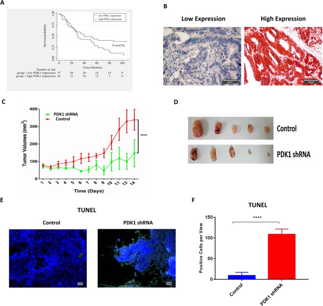
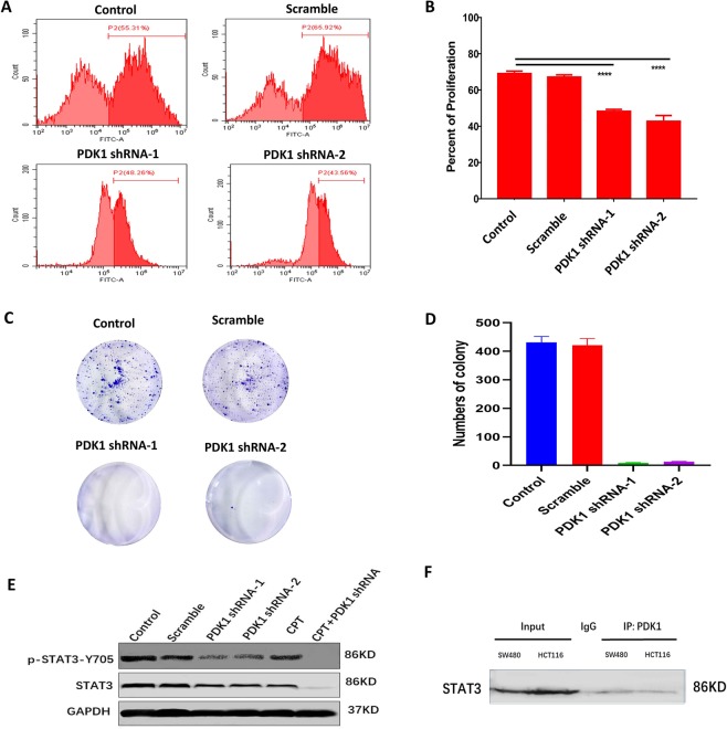
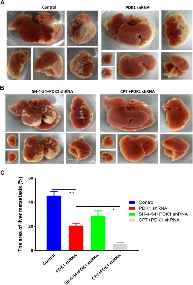
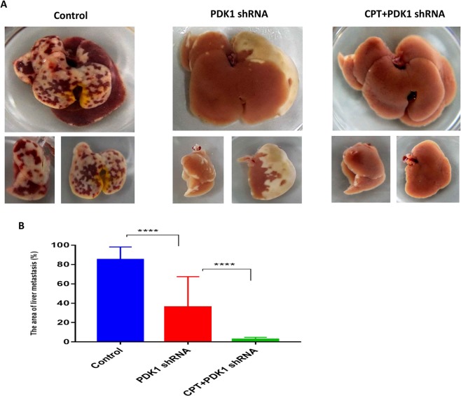
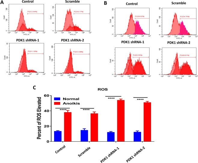
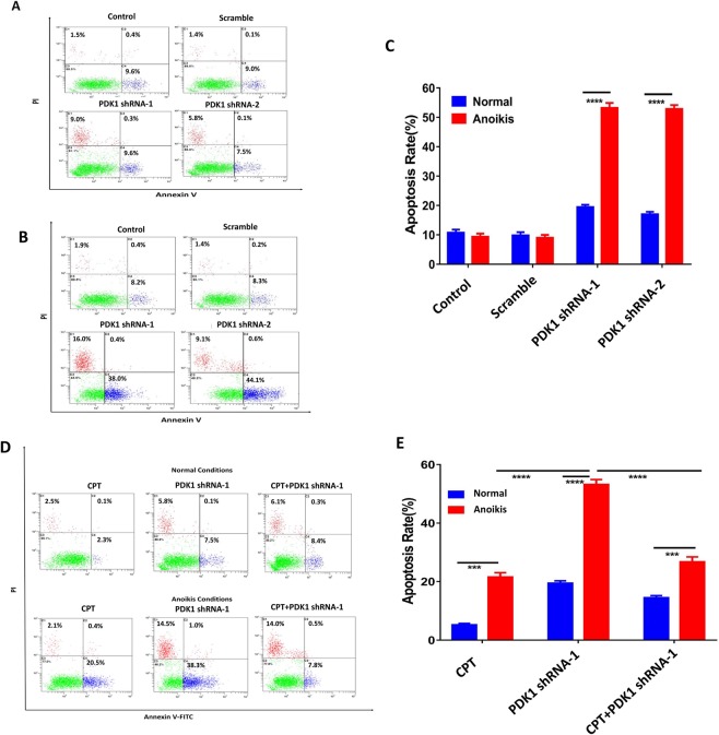
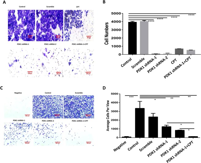
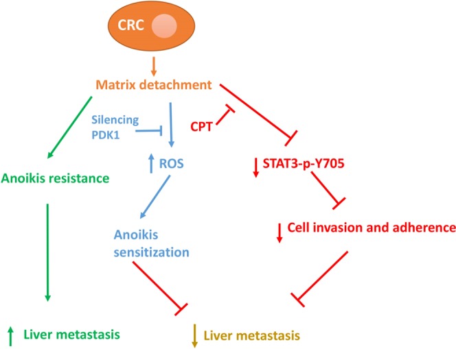
As a key glycolysis enzyme, the significance of pyruvate dehydrogenase kinase 1 (PDK1) in the development of colorectal cancer (CRC) remains unknown. This study revealed that the prognosis of CRC patients with high levels of PDK1 was poor, and PDK1 knockdown significantly reduced liver metastasis of CRC in both nude mice and immune competent BALB/C mice. When combined with cryptotanshinone (CPT), an inhibitor of STAT3-p-Y705, the liver metastasis was further inhibited. PDK1 knockdown obviously increased reactive oxygen species level in anoikis conditions and subsequently resulted in an elevated anoikis, but the combination of PDK1 knockdown and CPT showed a reduced effect on anoikis. Based on this discrepancy, the adherence ability of CRC cells to matrix protein fibronectin was further detected. It showed that PDK1 knockdown significantly decreased the adherence of CRC cells to fibronectin when combined with CPT. These results suggest that inhibition of PDK1 can decrease the surviving CRC cells in blood circulation via up-regulation of anoikis, and inhibition of STAT3-p-Y705 can prevent it to settle down on the liver premetastatic niche, which ultimately reduces liver metastasis.


Conflict of interest statement

The authors declare no competing interests.

Figures

Figure 1
Figure 1
High PDK1 expression is predictive of poor prognosis in CRC patients and promotes tumor growth in vivo. (A) Overall survival of CRC patients with high PDK1 expression (n = 42) was much shorter than patients with low PDK1 expression (n = 37). (B) IHC showed the representative results of low and high PDK1 expression based on the staining index on a tissue microarray. (C) Nude mice were subcutaneously injected with 8 × 106 HCT116 cells with or without the transduction of PDK1 shRNA. Silencing PDK1 significantly slowed down the growth of HCT116 xenografts (P < 0.0001). (D) The gross xenografts were isolated from each nude mouse. (E) TUNEL assay showed that silencing PDK1 promoted the apoptosis of CRC cells in residual xenograft of nude mice. (F) Histogram displayed the corresponding comparison of the apoptosis cells per view presented in (E). Data expressed as mean ± S.D., **** represents P < 0.0001.
Figure 2
Figure 2
The direct interaction between PDK1 and p-STAT3 may contribute to CRC proliferation. (A) EdU incorporation assay showed silencing PDK1 obviously decreased the proliferation of HCT116 cells in vitro, compared with the control. (B) Histogram displayed the corresponding comparison of the proliferation rate presented in (A). (C) Colony formation assay demonstrated that knockdown of PDK1 significantly decreased HCT116 cell colony formation in vitro. (D) Histogram illustrated the corresponding comparison of colony formation numbers presented in (C). (E) Western blot showed that knockdown of PDK1 significantly reduced STAT3-p-Y705 protein level in HCT116 cells. In particular, the Y705 phosphorylation was completely inhibited by the combination of CPT (a STAT3-p-Y705 inhibitor) and knockdown of PDK1. Three gels were loaded, and blots from different proteins were cropped and grouped into one image with white area separated in between different proteins. The exposure time was 50 s, 30 s, 10 s for p-STAT3, STAT3 and GAPDH, respectively. (F) A Co-IP showed PDK1 interacted directly with STAT3 in both HCT116 cells and SW480 cells. Rabbit IgG was served as the control. One gel was loaded, and the blot for STAT3 protein was cropped (exposure time: 40 s). Data expressed as mean ± S.D., **** represents P < 0.0001.
Figure 3
Figure 3
Knockdown of PDK1 and inhibiting p-STAT3-Y705 decreased liver metastasis of colon cancer in the nude mice model. (A) 5 × 106 HCT116 cells with or without PDK1 shRNA transduction were injected into the spleens of nude mice under isoflurane anesthesia on day 1, respectively. On day 9, the nude mice were sacrificed, and the liver metastatic area was measured by ImageJ software. Compared with the control group, knockdown of PDK1 significantly inhibited liver metastasis of HCT116 cells. (B) SH-4-54 (an inhibitor of total STAT3, 0.2 mg/d/mouse) or CPT (0.2 mg/d/mouse) were injected (i.p.) every other day (day 1, 3, 5, 7) for a total of four times. The results showed that CPT plus knockdown of PDK1 markedly decreased the liver metastasis area, compared with the combination of SH-4-54 and knockdown of PDK1. (C) Histogram indicated the corresponding comparison of liver metastasis areas presented in (A,B). Data expressed as mean ± S.D., * represents P < 0.05, ** represents P < 0.01.
Figure 4
Figure 4
The combination of CPT and silencing PDK1 significantly inhibited liver metastasis of CRC by down-regulating p-STAT3-Y705 in the immune competent mice model. (A) 5 × 106 HCT116 cells with or without PDK1 shRNA transduction were injected into the spleens of BALB/C mice under isoflurane anesthesia, respectively. CPT (0.2 mg/d/mouse) were injected (i.p.) every other day (day 1, 3, 5, 7) for a total of four times. On day 9, the mice were sacrificed, and the liver metastatic area was measured by ImageJ software. The combination of CPT and PDK1 shRNA treatment led to a smallest liver metastasis area, compared with the control or PDK1 shRNA treatment alone. (B) Histogram indicated the corresponding comparison of liver metastasis areas presented in (A). Data expressed as mean ± S.D., **** represents P < 0.0001.
Figure 5
Figure 5
Silencing PDK1 significantly elevates the cytoplasmic ROS level. (A) HCT116 cells with or without the transduction of a PDK1 shRNA were cultured in normal condition. Flow cytometry showed ROS levels had no obvious difference regardless of PDK1 status. (B) As indicated in (A), cells were cultured in matrix detachment. ROS levels were significantly enhanced by silencing PDK1, compared with the controls. (C) Histogram indicated the corresponding comparison of ROS levels presented in normal and anoikis culture conditions in (A,B). Data expressed as mean ± S.D., **** represents P < 0.0001.
Figure 6
Figure 6
Anoikis partly contributes to the in vivo effect of the combination of knockdown of PDK1 and CPT. (A) HCT116 cells with or without the transduction of a PDK1 shRNA were seeded into 6-well plates and cultured in normal conditions. Then the cells were stained with AnnexinV-FITC/PI. Flow cytometry showed that the apoptosis rate was much higher after PDK1 was silenced. (B) As indicated in (A), the cells were seeded into 6-well plates coated with poly-HEMA. Flow cytometry showed that the apoptotic rate of HCT116 cells stably transduced with PDK1 was significantly higher than the other two controls. (C) Histogram indicated the corresponding comparison of the apoptosis rate presented in (A,B). (D) The results showed knockdown of PDK1 strongly sensitized CRC to anoikis; however, the anoikis rate was significantly reduced when PDK1 silencing was combined with CPT. (E) Histogram indicated the corresponding comparison of the apoptosis results presented in both normal condition and suspending condition in (D). Data expressed as mean ± S.D., *** represents P < 0.001, and **** represents P < 0.0001.
Figure 7
Figure 7
Knockdown of PDK1 plus CPT significantly reduces liver metastasis in CRC by downregulating the adherence capacity via inhibiting STAT3-p-Y705. (A) Transwell assay showed that the knockdown of PDK1 or the combination of CPT and silencing PDK1 significantly decreased migration capacity of liver metastatic HCT116 cells. (B) Histogram indicated the corresponding comparison of migration capacity presented in (A). (C) The liver metastatic HCT116 cells were seeded into a 6-well plate coated with fibronectin. The adherence assay showed that the knockdown of PDK1 significantly decreased the chemotaxis of liver metastatic HCT116 cells. In particular, the combination of CPT and knockdown of PDK1 resulted in a lowest cell adherence capacity. (D) Histogram indicated the corresponding comparison of the adherent cells presented in (C). Data expressed as mean ± S.D., * represents P < 0.05, ** represents P < 0.01, *** represents P < 0.001, and **** represents P < 0.0001.
Figure 8
Figure 8
The potential mechanism of knockdown of PDK1 and CPT on inhibiting liver metastasis in CRC.

Similar articles

Cited by

References

    1. Ferlay J, et al. Cancer incidence and mortality worldwide: sources, methods and major patterns in GLOBOCAN 2012. Int J Cancer. 2015;136:E359–386. doi: 10.1002/ijc.29210. - DOI - PubMed
    1. Siegel RL, Miller KD, Jemal A. Cancer statistics, 2016. CA Cancer J Clin. 2016;66:7–30. doi: 10.3322/caac.21332. - DOI - PubMed
    1. House MG, et al. Comparison of adjuvant systemic chemotherapy with or without hepatic arterial infusional chemotherapy after hepatic resection for metastatic colorectal cancer. Ann Surg. 2011;254:851–856. doi: 10.1097/SLA.0b013e31822f4f88. - DOI - PubMed
    1. Tomlinson JS, et al. Actual 10-year survival after resection of colorectal liver metastases defines cure. J Clin Oncol. 2007;25:4575–4580. doi: 10.1200/JCO.2007.11.0833. - DOI - PubMed
    1. Jose C, Bellance N, Rossignol R. Choosing between glycolysis and oxidative phosphorylation: a tumor’s dilemma? Biochim Biophys Acta. 2011;1807:552–561. doi: 10.1016/j.bbabio.2010.10.012. - DOI - PubMed

Publication types

MeSH terms