

Patient-Specific Simulation of Pneumoperitoneum for Laparoscopic Surgical Planning
- PMID: 31506884
- PMCID: PMC6736924
- DOI: 10.1007/s10916-019-1441-z
Patient-Specific Simulation of Pneumoperitoneum for Laparoscopic Surgical Planning
Abstract
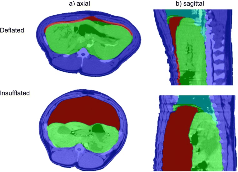
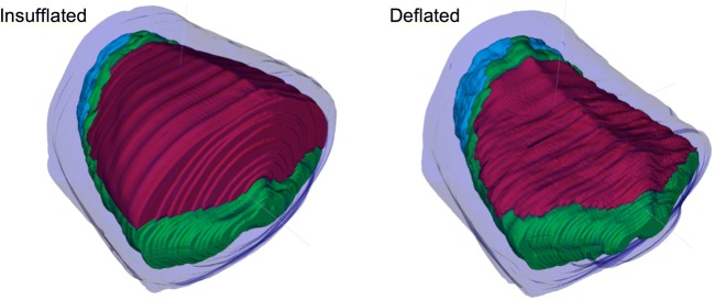
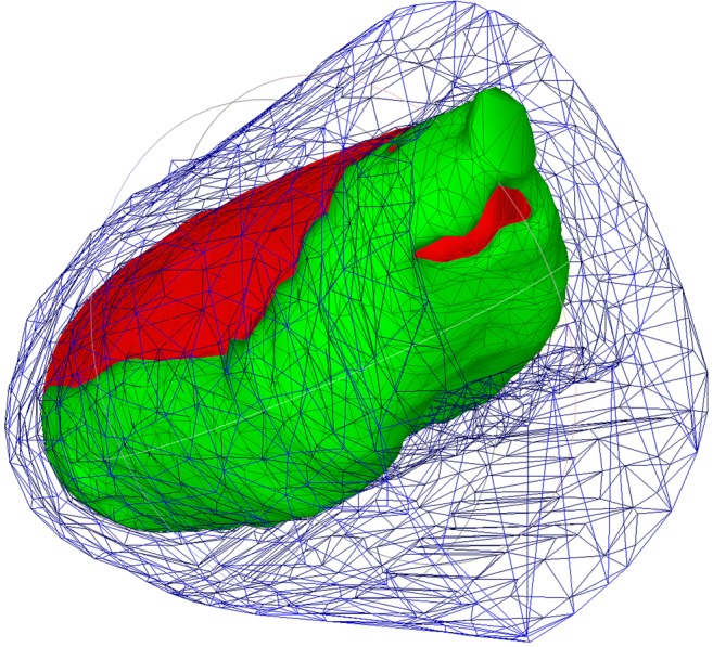
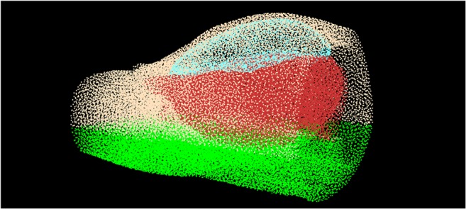
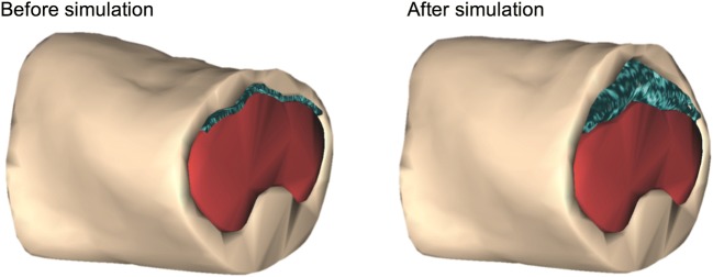
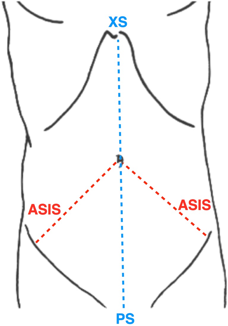
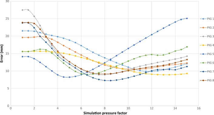
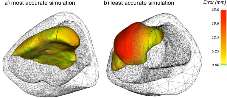
Gas insufflation in laparoscopy deforms the abdomen and stretches the overlying skin. This limits the use of surgical image-guidance technologies and challenges the appropriate placement of trocars, which influences the operative ease and potential quality of laparoscopic surgery. This work describes the development of a platform that simulates pneumoperitoneum in a patient-specific manner, using preoperative CT scans as input data. This aims to provide a more realistic representation of the intraoperative scenario and guide trocar positioning to optimize the ergonomics of laparoscopic instrumentation. The simulation was developed by generating 3D reconstructions of insufflated and deflated porcine CT scans and simulating an artificial pneumoperitoneum on the deflated model. Simulation parameters were optimized by minimizing the discrepancy between the simulated pneumoperitoneum and the ground truth model extracted from insufflated porcine scans. Insufflation modeling in humans was investigated by correlating the simulation's output to real post-insufflation measurements obtained from patients in theatre. The simulation returned an average error of 7.26 mm and 10.5 mm in the most and least accurate datasets respectively. In context of the initial discrepancy without simulation (23.8 mm and 19.6 mm), the methods proposed here provide a significantly improved picture of the intraoperative scenario. The framework was also demonstrated capable of simulating pneumoperitoneum in humans. This study proposes a method for realistically simulating pneumoperitoneum to achieve optimal ergonomics during laparoscopy. Although further studies to validate the simulation in humans are needed, there is the opportunity to provide a more realistic, interactive simulation platform for future image-guided minimally invasive surgery.
Keywords: Laparoscopy; Patient-specific; Pneumoperitoneum; Simulation; Surgical planning.
Conflict of interest statement
The authors declare that they have no conflict of interest.
Figures
Similar articles
-
Subject-specific modelling of pneumoperitoneum: model implementation, validation and human feasibility assessment.Int J Comput Assist Radiol Surg. 2019 May;14(5):841-850. doi: 10.1007/s11548-019-01924-2. Epub 2019 Feb 20. Int J Comput Assist Radiol Surg. 2019. PMID: 30788665 Free PMC article.
-
Simulation of pneumoperitoneum for laparoscopic surgery planning.Med Image Comput Comput Assist Interv. 2012;15(Pt 1):91-8. doi: 10.1007/978-3-642-33415-3_12. Med Image Comput Comput Assist Interv. 2012. PMID: 23285539
-
Comparison of a Valveless Trocar System and Conventional Insufflation in Pediatric Urologic Surgery.J Endourol. 2024 Jan;38(1):47-52. doi: 10.1089/end.2023.0181. Epub 2023 Nov 7. J Endourol. 2024. PMID: 37819689
-
Valveless Trocar Versus Standard Pneumoperitoneum Insufflation System in Minimally Invasive Surgery: Impact on Postoperative Pain. A Systematic Review and Meta-Analysis.J Laparoendosc Adv Surg Tech A. 2022 Sep;32(9):978-986. doi: 10.1089/lap.2022.0022. Epub 2022 Apr 11. J Laparoendosc Adv Surg Tech A. 2022. PMID: 35404130
-
Laparoscopic entry: a review of techniques, technologies, and complications.J Obstet Gynaecol Can. 2007 May;29(5):433-447. doi: 10.1016/S1701-2163(16)35496-2. J Obstet Gynaecol Can. 2007. PMID: 17493376 Review. English, French.
Cited by
-
The technology of artificial pneumoperitoneum CT and its application in diagnosis of abdominal adhesion.Sci Rep. 2021 Oct 21;11(1):20785. doi: 10.1038/s41598-021-00408-1. Sci Rep. 2021. PMID: 34675300 Free PMC article.
-
Initial Report: A Novel Intraoperative Navigation System for Laparoscopic Liver Resection Using Real-Time Virtual Sonography.Sci Rep. 2020 Apr 10;10(1):6174. doi: 10.1038/s41598-020-63131-3. Sci Rep. 2020. PMID: 32277107 Free PMC article.
References
-
- Kitasaka Takayuki, Mori Kensaku, Hayashi Yuichiro, Suenaga Yasuhito, Hashizume Makoto, Toriwaki Jun-ichiro. Medical Image Computing and Computer-Assisted Intervention – MICCAI 2004. Berlin, Heidelberg: Springer Berlin Heidelberg; 2004. Virtual Pneumoperitoneum for Generating Virtual Laparoscopic Views Based on Volumetric Deformation; pp. 559–567.
-
- Bano, J., Hostettler, A., Nicolau, S. A., Cotin, S., Doignon, C., Wu, H. S. et al., Simulation of pneumoperitoneum for laparoscopic surgery planning. Med. Image Comput. Comput. Interv. – MICCAI 2012, 2012, 91–98. - PubMed
MeSH terms
Grants and funding
LinkOut - more resources
Full Text Sources
Medical
