Review
. 2020 Jan;97(1):138-155.
doi: 10.1111/cge.13641. Epub 2019 Oct 23.

Growth factor and receptor malfunctions associated with human genetic deafness

Affiliations
Review

Growth factor and receptor malfunctions associated with human genetic deafness

Sadaf Naz et al. Clin Genet. 2020 Jan.

Abstract

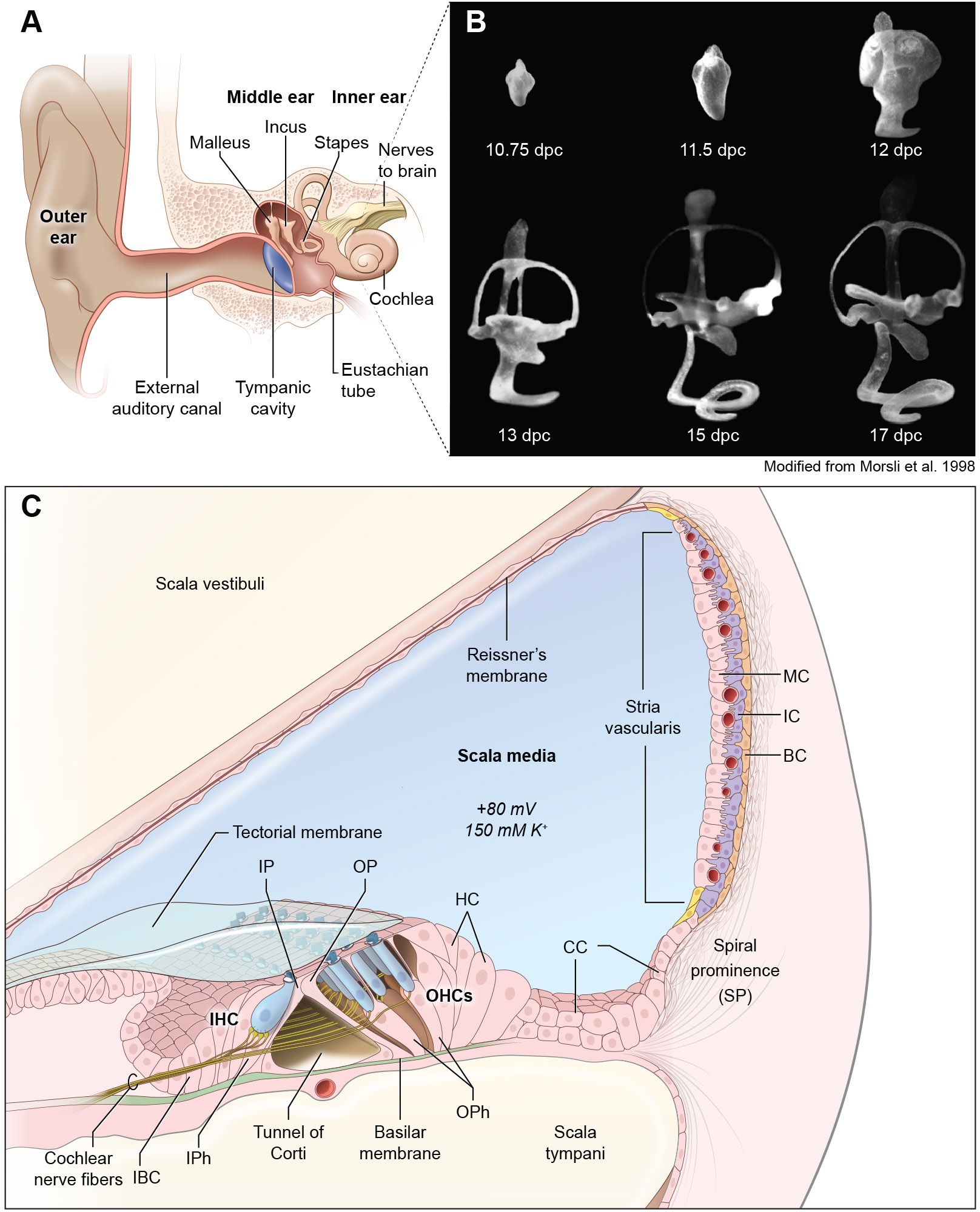
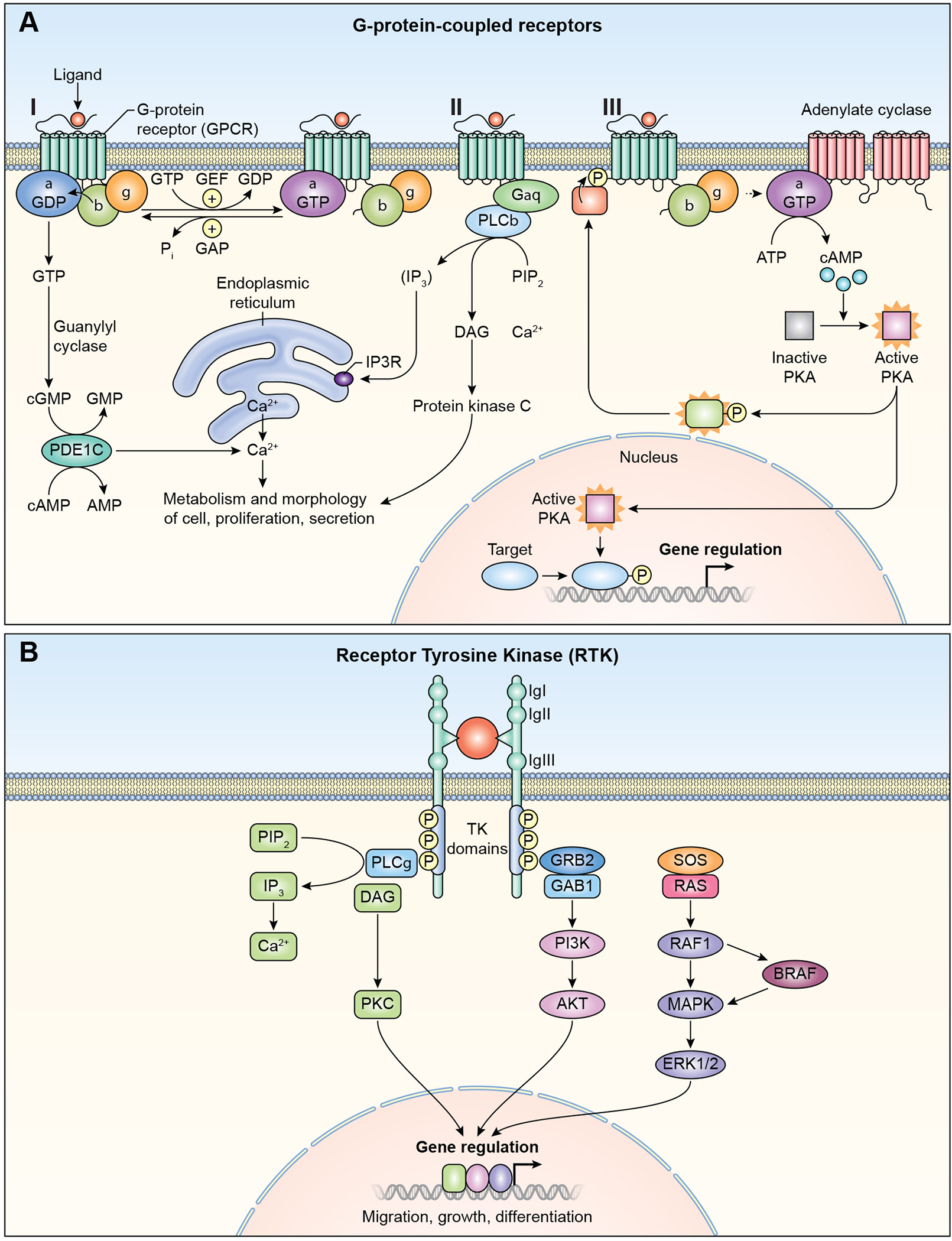
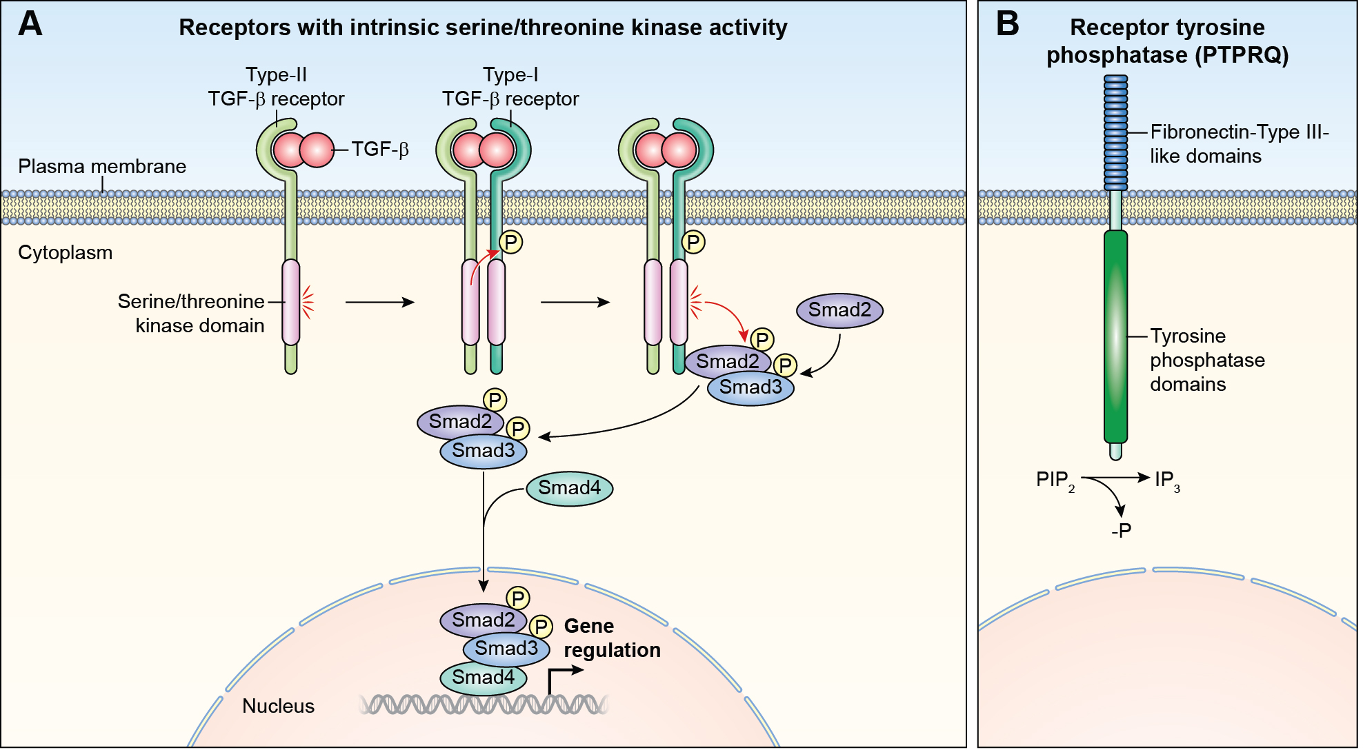
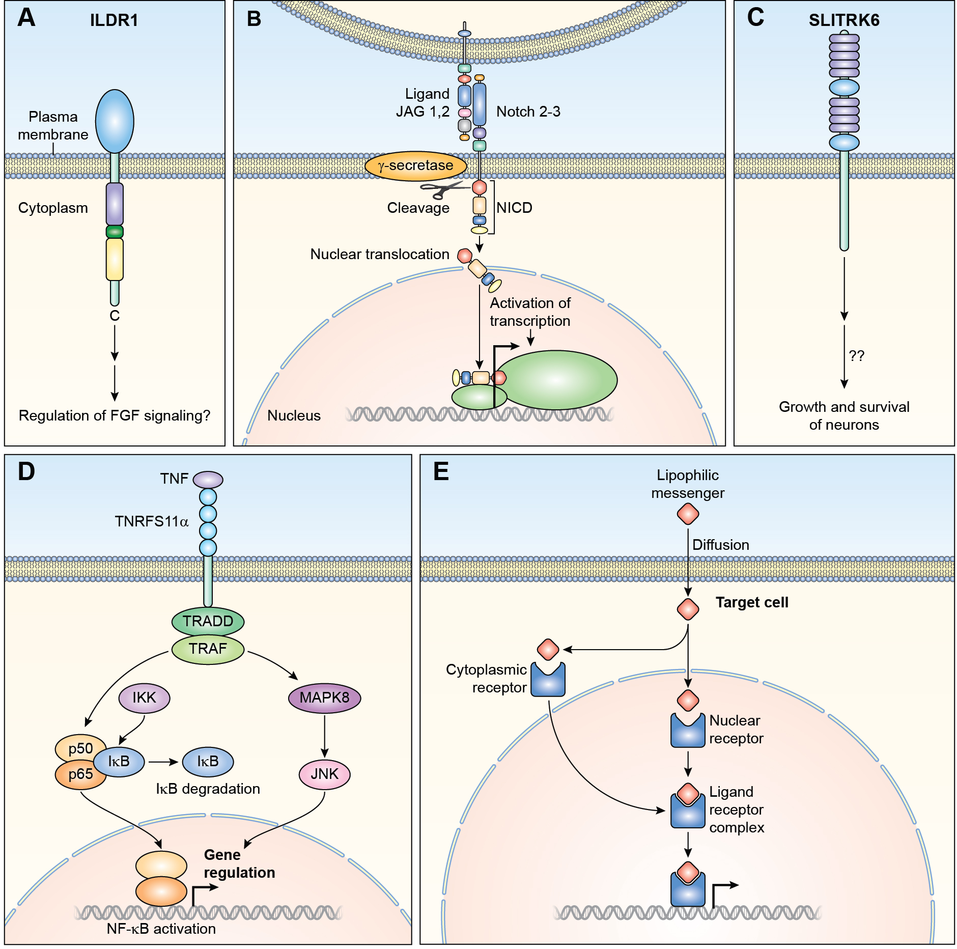
A variety of different signaling pathways are necessary for development and maintenance of the human auditory system. Normal hearing allows for the detection of soft sounds within the frequency range of 20 to 20 000 Hz, but more importantly to perceive the human voice frequency band of 250 to 6000 Hz. Loss of hearing is common, and is a clinically heterogeneous disorder that can be caused by environmental factors such as exposure to loud noise, infections and ototoxic drugs. In addition, variants of hundreds of genes have been reported to disrupt processes required for hearing. Noncoding regulatory variants and variants of additional genes necessary for hearing remain to be discovered as many individuals with inherited deafness are without a genetic diagnosis, despite the advent of whole exome sequencing. Here, we discuss in detail some of these deafness-causing variants of genes encoding a ligand or its receptor. Spotlighted in this review are three growth factor-receptor-pairs EDN3/EDNRB, HGF/MET and JAG/NOTCH, which individually are necessary for normal hearing. We also offer our perspective on unanswered questions, future challenges and potential opportunities for treatments emerging from molecular genetic and mechanistic studies of deafness due to these causes.

Keywords: EDN3; EDNRB; HGF; JAG; MET; NOTCH; hearing; inner ear defects.


Conflict of interest statement

DISCLOSURE

The authors declare no conflict of interest

Figures

FIGURE 1.
FIGURE 1.
Development and structure of the ear. (A) Structure of the human ear showing its three main parts, outer, middle and inner ear. (B) Paint-filled mouse membranous labyrinths at embryonic days 10.75 days-postcoitum to postnatal day 1 (P1). Lateral views are shown. Scale bar, 200 μm. (C) Diagram of a cross-section of the cochlea. The roof of the cochlear duct is formed by two layers of flattened cells comprising Reissner’s membrane, while the base is formed by the basilar membrane, which separate the cochlear duct (scala media) from the scala vestibuli and the scala tympani. The three rows of outer hair cells, one row of inner hair cells and different types of supporting cells and the stria vascularis are shown. IHC; Inner Hair Cells, OHC, Outer Hair Cells, IP, Inner Pillar cells, OP; Outer Pillar Cells, HC; Hensen’s Cells, CC; Claudius Cells, BC; Basal Cells, IC; Intermediate cells, MC; Marginal Cells, IBC; inner border cells.
FIGURE 2.
FIGURE 2.
GPCR and RTK signaling pathways. (A) G-protein Coupled Receptors (GPCR). I. The receptors are coupled to the C-terminus of heterotrimeric G-proteins. G-proteins are composed of α, β, and γ subunits. GPCRs activate signaling through multiple pathways. II. Phospholipase C, (PLC), phosphatidylinositol signaling pathway. Diacyl glycerol (DAG) and inositol 1,4,5-trisphosphate (IP3) are generated by cleavage of phospholipid phosphatidylinositol 4,5-bisphosphate (PIP2) by phospholipase C. IP3 releases calcium ions from endoplasmic reticulum. Diacylglycrol remains bound to the plasma membrane. Both diacylglycerol and calcium ions act together activate Protein Kinase C (PKC). PKC phosphorylates multiple cytoplasmic proteins that regulate cellular activity. III. Cyclic AMP signaling pathway. Cyclic AMP is a second messenger. GPCR activates adenylate cyclase, which converts ATP to cyclic AMP (cAMP). cAMP activates protein Kinase A (PKA), which then phosphorylates different proteins and transcription factors. PKA translocates to nucleus and controls gene transcription of target genes. IP3R, inositol trisphoshphate receptor, GEF; guanidine exchange factor, P; Phosphate. (B) Receptor Tyrosine Kinase (RTK). Ligand binding causes receptor dimerization and autophosphorylation. Signaling via RTK can take place through phospholipase C pathway, left side of figure (also see Figure 2A). GRB2 with other associated proteins such as GAB1, is bound to RTK. The activated receptor phosphorylates SOS1 and other guanine nucleotide exchange factors (GEF), and members of RAS, RHO and RAF, which are tethered to membranes. Signals are further propagated through the MAPK/ERK pathway to regulate gene expression. PI3K; phosphoinositide 3- kinase, Akt; Protein Kinase B.
FIGURE 3.
FIGURE 3.
Other receptors with intrinsic enzymatic activity (A) Receptor serine/threonine kinase. A ligand of the TGFB superfamily or the BMP superfamily, binds first to a type II receptor, which in turn phosphorylates the type I receptor and leads to their dimerization. The activated receptor complex phosphorylates the SMAD family proteins. Phosphorylation of SMAD proteins disassociates them from the receptor complex. Bound SMADS translocate to the nucleus and form complexes with DNA regulatory proteins inhibiting or activating transcription of target genes. (B) Receptor tyrosine phosphatase. PTPRQ dephosphorylates phosphatidylinositol 3,4,5-trisphosphate (PIP3) to PIP2. (See Figure 2B for details of signaling via PIP2).
FIGURE 4.
FIGURE 4.
Non-enzymatic receptors (A) ILDR1 has an extracellular immunoglobin like domain and intracellular cysteine and arginine regions. Both of these are separated by a dileucine motif. Signaling through this receptor in the inner ear remains to be elucidated. (B) NOTCH receptors bind to transmembrane ligands such as JAG1. As a result of binding, NOTCH is proteolytically cleaved by γ-secretase releasing the intracellular domain of the protein (NICD), which translocates to the nucleus forming complexes with other proteins and regulate transcription of target genes. (C) SLITRK6 is a transmembrane receptor with leucine rich repeats in its extracellular domain. The mechanism of signaling through SLITRK6 is unknown. (D) TRADD and TRAFF are adapters of TNRFS receptors and participate in downstream signaling. Binding of TNF to its receptors activates either MAPK8/JNK or NF-kB (NFKB1), which is sequestered in the cytoplasm by IKB. The activation of NFKB1 by receptor binding activates a kinase (IKK) which phosphorylates IKB at specific serine residues. IKB is then ubiquitinated and degraded by the proteasome. Free NFKB1 translocates to the nucleus and regulates transcription. The second signaling pathway involves MAPK8/JNK. MAPK8 phosphorylates a number of transcription factors which modulate transcription of specific genes. (E) Ligand binding to nuclear hormone receptors cause dimerization and translocation to the nucleus. Receptor-DNA binding controls transcription of targeted genes involved in a variety of cellular activities including ion transport and proliferation. IKB; nuclear factor of kappa light polypeptide gene enhancer in B-cells inhibitor, alpha, IKK; IKB Kinase.

References

    1. Tucci D, Merson MH, Wilson BS. A summary of the literature on global hearing impairment: current status and priorities for action. Otol Neurotol. 2010;31(1):31–41. - PubMed
    1. Yueh B, Shapiro N, MacLean CH, Shekelle PG. Screening and management of adult hearing loss in primary care: scientific review. JAMA. 2003;289(15):1976–1985. - PubMed
    1. Fritzsch B, Elliott KL. Gene, cell, and organ multiplication drives inner ear evolution. Dev Biol. 2017;431(1):3–15. - PMC - PubMed
    1. Morsli H, Choo D, Ryan A, Johnson R, Wu DK. Development of the mouse inner ear and origin of its sensory organs. J Neurosci. 1998;18(9):3327–3335. - PMC - PubMed
    1. Purves D, Augustine GJ, Fitzpatrick D, et al., eds. Neuroscience. 2 ed: Sunderland; 2001.

Publication types

MeSH terms