

Solitary Pulmonary Nodule: Morphological Effects on Metabolic Activity Assessment
- PMID: 31507144
- PMCID: PMC6746010
- DOI: 10.4274/mirt.galenos.2019.65707
Solitary Pulmonary Nodule: Morphological Effects on Metabolic Activity Assessment
Abstract
Objectives: We aimed to evaluate the effects of morphological characteristics of the solitary pulmonary nodules (SPN) on metabolic activity assessment. To the best of our knowledge, this is the first study to compare the volumetric metabolic activity parameters according to the morphologic parameters of the nodules.
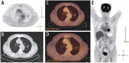
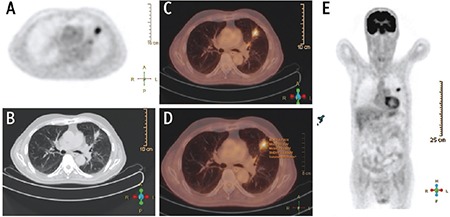
Methods: In this retrospective study, 18F-FDG positron emission tomography and computed tomography scans performed between 2011 and 2018 were evaluated by a nuclear and diagnostic radiologist. One hundred thirteen patients with SPNs with biopsy-proven diagnosis were included. SPNs were classified as solid, partially solid (PS), and ground glass opacity (GGO).
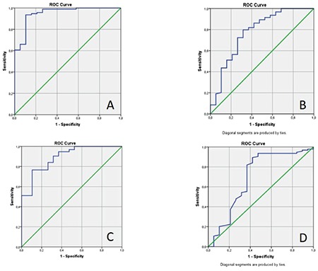
Results: SPN diameter, SUVmax, metabolic tumor volume (MTV), total lesion glycolysis (TLG), and density were significantly higher in the malignant group. SUVmax, MTV, TLG increased in direct proportion to the diameter. There was no a significant difference between GGO, PS, and solid nodules in terms of SUVmax values. MTV and TLG values increased in parallel with the density of the nodules, but this increase was only significant in the malignant group. There was a significant difference between SPNs <2 cm and SPNs ≥2 cm in terms of MTV, while there was no difference in terms of SUVmax. The cut-off value determined by the ROC curve was found to be 4.39 for SUVmax, 7.33 mL for MTV and 31.88 g for TLG. The cut-off values for SUVmax of solid and subsolid nodules were close to each other, but cut-off values for MTV and TLG were higher in solid nodules.
Conclusion: SUVmax, MTV, and TLG are affected by diameter and attenuation. We suggest using different MTV and TLG cut-off values for solid and subsolid nodules, but we suggest using same SUVmax values. MTV can be a more reliable parameter than SUVmax in prediction of malignancy in smaller nodules.
Amaç: Soliter pulmoner nodüllerin (SPN) morfolojik özelliklerinin metabolik aktivite değerlendirmesi üzerindeki etkilerini değerlendirmeyi amaçladık. Bildiğimiz kadarıyla, volümetrik metabolik aktivite parametrelerini nodüllerin morfolojik parametrelerine göre karşılaştıran ilk çalışma budur.
Yöntem: 2011 ve 2018 yılları arasında yapılan 18F-FDG pozitron emisyon tomografisi ve bilgisayarlı tomografi taramaları, bir nükleer tıp uzmanı ve radyoloji uzmanı tarafından retrospektif olarak değerlendirildi. Biyopsi ile kanıtlanmış tanısı olan 113 SPN hastası dahil edildi. SPN’ler solid, kısmi solid (PS) ve buzlu cam opasitesi (GGO) olarak sınıflandırıldı
Bulgular: SPN çapı, SUVmaks, metabolik tümör hacmi (MTV), toplam lezyon glikoliz (TLG) ve dansite malign grupta anlamlı olarak yüksek bulundu. SUVmaks, MTV ve TLG değerleri çap ile doğru orantılı olarak arttı. GGO, PS ve solid nodüller arasında SUVmaks değerleri arasında anlamlı bir fark yoktu. MTV, TLG değerleri nodüllerin yoğunluğu ile paralel olarak arttı; ancak sadece malign grupta anlamlı fark bulundu. 2 cm’den küçük grupta MTV için anlamlı fark varken SUVmaks için yoktu. ROC eğrisi ile belirlenen kesme değerinin SUVmaks için 4,39, MTV için 7,33 mL, TLG için 31,88 g olduğu bulundu. Solid ve subsolid nodüllerin SUVmaks için cut-off değeri birbirine yakındı, ancak MTV, TLG için cut-off değer solid nodüllerde daha yüksekti.
Sonuç: SUVmaks, MTV ve TLG çap ve atenüasyondan etkilenmektedir. Solid ve subsolid nodüller için farklı MTV ve TLG cut-off değerlerinin kullanılmasının gerektiğini; ancak SUVmaks için gerekli olmadığını düşünmekteyiz. MTV, küçük nodüller için malignite tahmininde SUVmaks’tan daha güvenilir bir parametre olabilir.
Conflict of interest statement
Figures
Similar articles
-
Qualitative and Semiquantitative Parameters of 18F-FDG-PET/CT as Predictors of Malignancy in Patients with Solitary Pulmonary Nodule.Cancers (Basel). 2023 Feb 4;15(4):1000. doi: 10.3390/cancers15041000. Cancers (Basel). 2023. PMID: 36831344 Free PMC article.
-
Assessment of Mediastinal Tumors Using SUVmax and Volumetric Parameters on FDG-PET/CT.Asia Ocean J Nucl Med Biol. 2017 Winter;5(1):22-29. doi: 10.22038/aojnmb.2016.7996. Asia Ocean J Nucl Med Biol. 2017. PMID: 28840135 Free PMC article.
-
Prognostic Value of Volumetric Parameters Measured by Pretreatment 18F FDG PET/CT in Patients With Cutaneous Malignant Melanoma.Clin Nucl Med. 2016 Jun;41(6):e266-73. doi: 10.1097/RLU.0000000000001205. Clin Nucl Med. 2016. PMID: 27055144
-
Prognostic Value of 18F-FDG PET/CT in Surgical Non-Small Cell Lung Cancer: A Meta-Analysis.PLoS One. 2016 Jan 4;11(1):e0146195. doi: 10.1371/journal.pone.0146195. eCollection 2016. PLoS One. 2016. PMID: 26727114 Free PMC article. Review.
-
Prognostic value of maximum standard uptake value, metabolic tumor volume, and total lesion glycolysis of positron emission tomography/computed tomography in patients with breast cancer: A systematic review and meta-analysis.PLoS One. 2019 Dec 11;14(12):e0225959. doi: 10.1371/journal.pone.0225959. eCollection 2019. PLoS One. 2019. PMID: 31826010 Free PMC article.
Cited by
-
Amide proton transfer-weighted imaging and stretch-exponential model DWI based 18F-FDG PET/MRI for differentiation of benign and malignant solitary pulmonary lesions.Cancer Imaging. 2024 Mar 4;24(1):33. doi: 10.1186/s40644-024-00677-9. Cancer Imaging. 2024. PMID: 38439101 Free PMC article.
-
PET/CT-aided biopsy of lung lesions enhances diagnostic efficacy, especially for lesions >3cm.Front Oncol. 2024 Jan 30;14:1296553. doi: 10.3389/fonc.2024.1296553. eCollection 2024. Front Oncol. 2024. PMID: 38357204 Free PMC article.
-
Predicting solitary pulmonary lesions in breast cancer patients using 18fluorodeoxyglucose-positron emission tomography/computed tomography combined with clinicopathological characteristics.BMC Pulm Med. 2024 Nov 29;24(1):595. doi: 10.1186/s12890-024-03418-7. BMC Pulm Med. 2024. PMID: 39614273 Free PMC article.
-
Qualitative and Semiquantitative Parameters of 18F-FDG-PET/CT as Predictors of Malignancy in Patients with Solitary Pulmonary Nodule.Cancers (Basel). 2023 Feb 4;15(4):1000. doi: 10.3390/cancers15041000. Cancers (Basel). 2023. PMID: 36831344 Free PMC article.
References
-
- Tuddenham WJ. Glossary of terms for thoracic radiology: recommendations of the Nomenclature Committee of the Fleischner Society. AJR Am J Roentgenol. 1984;143:509–517. - PubMed
-
- Ost D, Fein AM, Feinsilver SH. Clinical practice. The solitary pulmonary nodule. N Engl J Med. 2003;348:2535–2542. - PubMed
-
- Midthun DE, Swensen SJj, Jett JR. Approach to the solitary pulmonary nodule. Mayo Clin Proc. 1993;68:378–385. - PubMed
-
- Gohagan J, Marcus P, Fagerstrom R, Pinsky P, Kramer B, Prorok P; Writing Committee, Lung Screening Study Research Group. Baseline findings of a randomized feasibility trial of lung cancer screening with spiral CT scan vs chest radiograph: the Lung Screening Study of the National Cancer Institute. Chest. 2004;126:114–121. - PubMed
-
- Swensen SJ, Jett JR, Hartman TE, Midthun DE, Sloan JA, Sykes AM, Aughenbaugh GL, Clemens MA. Lung cancer screening with CT: Mayo Clinic experience. Radiology. 2003;226:756–761. - PubMed
LinkOut - more resources
Full Text Sources