Review
. 2019 Sep;32(5):372-376.
doi: 10.1055/s-0039-1687833. Epub 2019 Sep 6.

Systemic and Autoimmune Diseases

Affiliations
Review

Systemic and Autoimmune Diseases

Xavier Delgadillo et al. Clin Colon Rectal Surg. 2019 Sep.

Abstract

This article reviews the clinical features of systemic and autoimmune diseases affecting the perianal region and its surrounding integumentary structures.

Keywords: acanthosis nigricans; glucagonoma syndrome; leukemic infiltration; pemphigus vulgaris; psoriasis; pyoderma gangrenosum; vitiligo.


Conflict of interest statement

Conflict of Interest None.

Figures

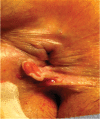
Fig. 1
Fig. 1
Clinical presentation of anal pemphigus vulgaris. A pseudofissure is present at the anal verge. Pain is usually present spontaneously or with defecation. (This image is provided courtesy of A. Ortega.)
Fig. 2
Fig. 2
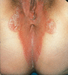
Pyoderma gangrenosum showing the irregular, violaceous, and undermined border surrounding the deep ulcer on the gluteal areas. (This image is provided courtesy of H. Foong.)
Fig. 3
Fig. 3
Perianal psoriasis. Red well-demarked plaques are evident. White scales are present in some parts of the dermatoses, while others are reddish and macerated. (This image is provided courtesy of X. Delgadillo.)
Fig. 4
Fig. 4
Acanthosis nigricans extending from the gluteal cleft to the thighs. (This image is provided courtesy of X. Delgadillo.)
Fig. 5
Fig. 5
Lesions spread peripherally with central desquamation and pallor, and associated circinate patterns are associated with perianal glucagonoma syndrome. (This image is provided courtesy of E. Csatar.)
Fig. 6
Fig. 6
Leukemic cutis ani infiltration. (This image is provided courtesy of E. Csatar.)
Fig. 7
Fig. 7
Perianal vitiligo in a noninflammatory bowel disease and asymptomatic patient. (This image is provided courtesy of A. Ortega.)

References

    1. Khezri S, Mahmoudi H R, Masoom S N et al.Anal involvement in pemphigus vularis. Autoimmune Dis. 2013;2013:609181. - PMC - PubMed
    1. Madala J, Bashamalla R, Kumar M P. Current concepts of pemphigus with a deep insight into its molecular aspects. J Oral Maxillofac Pathol. 2017;21(02):260–263. - PMC - PubMed
    1. Bystryn J C, Grando S A. A novel explanation for acantholysis in pemphigus vulgaris: the basal cell shrinkage hypothesis. J Am Acad Dermatol. 2006;54(03):513–516. - PubMed
    1. Malik M, El Tal A E, Ahmed A R. Anal involvement in pemphigus vulgaris. Dis Colon Rectum. 2006;49(04):500–506. - PubMed
    1. Vallini V, Andreini R, Bonadio A. Pyoderma gangrenosum: a current problem as much as an unknown one. Int J Low Extrem Wounds. 2017;16(03):191–201. - PubMed