

Early Pain Exposure Influences Functional Brain Connectivity in Very Preterm Neonates
- PMID: 31507370
- PMCID: PMC6716476
- DOI: 10.3389/fnins.2019.00899
Early Pain Exposure Influences Functional Brain Connectivity in Very Preterm Neonates
Abstract
Background: Early exposure to nociceptive events may cause brain structural alterations in preterm neonates, with long-lasting consequences on neurodevelopmental outcome. Little is known on the extent to which early pain may affect brain connectivity. We aim to evaluate brain functional connectivity changes in preterm neonate that underwent multiple invasive procedures during the postnatal period, and to correlate them with the neurodevelopmental outcome at 24 months.
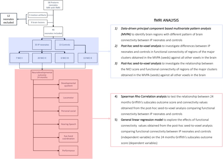
Methods: In this prospective case-control study, we collected information about exposure to painful events during the early postnatal period and resting-state BOLD-fMRI data at term equivalent age from two groups of preterm neonate: 33 subjected to painful procedures during the neonatal intensive care (mean gestational age 27.9 ± 1.8 weeks) and 13 who did not require invasive procedures (average gestational age 31.2 ± 2.1 weeks). A data-driven principal-component-based multivariate pattern analysis (MVPA) was used to investigate the effect of early pain exposure on brain functional connectivity, and the relationship between connectivity changes and neurodevelopmental outcome at 24 months, assessed with Griffiths, Developmental Scale-Revised: 0-2.
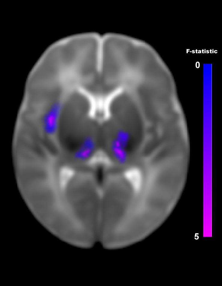
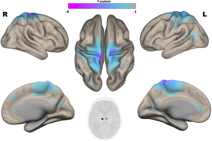
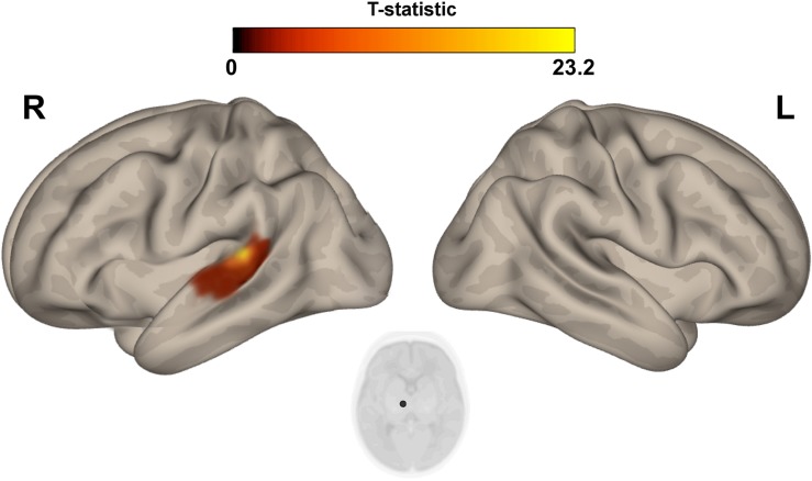
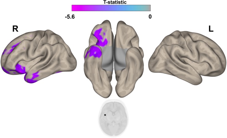
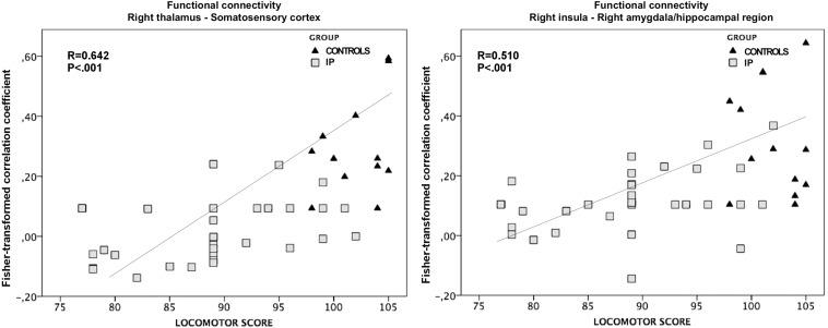
Results: Early pain was associated with decreased functional connectivity between thalami and bilateral somatosensory cortex, and between the right insular cortex and ipsilateral amygdala and hippocampal regions, with a more evident effect in preterm neonate undergoing more invasive procedures. Functional connectivity of the right thalamocortical pathway was related to neuromotor outcome at 24 months (P = 0.003).
Conclusion: Early exposure to pain is associated with abnormal functional connectivity of developing networks involved in the modulation of noxious stimuli in preterm neonate, contributing to the neurodevelopmental consequence of preterm birth.
Keywords: brain connectivity; fMRI; functional connectivity; neonatal neuroimaging; nociceptive modulations; pain; preterm neonates; resting state.
Figures
Similar articles
-
Early Procedural Pain Is Associated with Regionally-Specific Alterations in Thalamic Development in Preterm Neonates.J Neurosci. 2018 Jan 24;38(4):878-886. doi: 10.1523/JNEUROSCI.0867-17.2017. Epub 2017 Dec 18. J Neurosci. 2018. PMID: 29255007 Free PMC article.
-
Hippocampus, Amygdala, and Thalamus Volumes in Very Preterm Children at 8 Years: Neonatal Pain and Genetic Variation.Front Behav Neurosci. 2019 Mar 19;13:51. doi: 10.3389/fnbeh.2019.00051. eCollection 2019. Front Behav Neurosci. 2019. PMID: 30941021 Free PMC article.
-
Breastfeeding improves dynamic reorganization of functional connectivity in preterm infants: a temporal brain network study.Med Biol Eng Comput. 2020 Nov;58(11):2805-2819. doi: 10.1007/s11517-020-02244-3. Epub 2020 Sep 18. Med Biol Eng Comput. 2020. PMID: 32945999
-
Biological and neurodevelopmental implications of neonatal pain.Clin Perinatol. 2013 Sep;40(3):471-91. doi: 10.1016/j.clp.2013.05.002. Epub 2013 Jul 3. Clin Perinatol. 2013. PMID: 23972752 Review.
-
The impact of pain in the immature brain.J Matern Fetal Neonatal Med. 2009 Sep;22(9):722-32. doi: 10.3109/14767050902926962. J Matern Fetal Neonatal Med. 2009. PMID: 19526425 Review.
Cited by
-
Neonatal amygdala volumes, procedural pain and the association with social-emotional development in children born very preterm.Brain Struct Funct. 2024 Dec;229(9):2369-2378. doi: 10.1007/s00429-024-02845-w. Epub 2024 Aug 6. Brain Struct Funct. 2024. PMID: 39103553
-
Pain management for necrotizing enterocolitis: getting the balance right.Pediatr Res. 2022 Nov;92(5):1423-1431. doi: 10.1038/s41390-022-01968-2. Epub 2022 Feb 15. Pediatr Res. 2022. PMID: 35169278 Free PMC article.
-
Functional connectivity of the pediatric brain.Neuroradiology. 2024 Nov;66(11):2071-2082. doi: 10.1007/s00234-024-03453-5. Epub 2024 Sep 4. Neuroradiology. 2024. PMID: 39230715 Review.
-
Pain Exposure and Brain Connectivity in Preterm Infants.JAMA Netw Open. 2024 Mar 4;7(3):e242551. doi: 10.1001/jamanetworkopen.2024.2551. JAMA Netw Open. 2024. PMID: 38488791 Free PMC article.
-
Terminology matters: is the International Association for the Study of Pain definition of pain fully satisfactory for fetuses, neonates, and infants?Front Pain Res (Lausanne). 2024 May 16;5:1369945. doi: 10.3389/fpain.2024.1369945. eCollection 2024. Front Pain Res (Lausanne). 2024. PMID: 38818234 Free PMC article. No abstract available.
References
LinkOut - more resources
Full Text Sources