Altered Functional Connectivity in Patients With Sloping Sensorineural Hearing Loss
- PMID: 31507391
- PMCID: PMC6713935
- DOI: 10.3389/fnhum.2019.00284
Altered Functional Connectivity in Patients With Sloping Sensorineural Hearing Loss
Abstract
Background: Sensory deprivation, such as hearing loss, has been demonstrated to change the intrinsic functional connectivity (FC) of the brain, as measured with resting-state functional magnetic resonance imaging (rs-fMRI). Patients with sloping sensorineural hearing loss (SNHL) are a unique population among the hearing impaired, as they have all been exposed to some auditory input throughout their lifespan and all use spoken language.
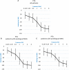
Materials and methods: Twenty patients with SNHL and 21 control subjects participated in a rs-fMRI study. Whole-brain seed-driven FC maps were obtained, with audiological scores of patients, including hearing loss severity and speech performance, used as covariates.
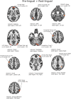
Results: Most profound differences in FC were found between patients with prelingual (before language development, PRE) vs. postlingual onset (after language development, POST) of SNHL. An early onset was related to enhancement in long-range network connections, including the default-mode network, the dorsal-attention network and the fronto-parietal network, as well as in local sensory networks, the visual and the sensorimotor. A number of multisensory brain regions in frontal and parietal cortices, as well as the cerebellum, were also more internally connected. We interpret these effects as top-down mechanisms serving optimization of multisensory experience in SNHL with a prelingual onset. At the same time, POST patients showed enhanced FC between the salience network and multisensory parietal areas, as well as with the hippocampus, when they were compared to those with PRE hearing loss. Signal in several cortex regions subserving visual processing was also more intra-correlated in POST vs. PRE patients. This outcome might point to more attention resources directed to multisensory as well as memory experience. Finally, audiological scores correlated with FC in several sensory and high-order brain regions in all patients.
Conclusion: The results show that a sloping hearing loss is related to altered resting-state brain organization. Effects were shown in attention and cognitive control networks, as well as visual and sensorimotor regions. Specifically, we found that even in a partial hearing deficit (affecting only some of the hearing frequency ranges), the age at the onset affects the brain function differently, pointing to the role of sensitive periods in brain development.
Keywords: functional connectivity; neuronal plasticity; partial deafness; resting state; sensorineural hearing loss.
Figures


Similar articles
-
Prefrontal-Temporal Pathway Mediates the Cross-Modal and Cognitive Reorganization in Sensorineural Hearing Loss With or Without Tinnitus: A Multimodal MRI Study.Front Neurosci. 2019 Mar 12;13:222. doi: 10.3389/fnins.2019.00222. eCollection 2019. Front Neurosci. 2019. PMID: 30930739 Free PMC article.
-
Altered resting-state functional network connectivity in profound sensorineural hearing loss infants within an early sensitive period: A group ICA study.Hum Brain Mapp. 2021 Sep;42(13):4314-4326. doi: 10.1002/hbm.25548. Epub 2021 Jun 1. Hum Brain Mapp. 2021. PMID: 34060682 Free PMC article.
-
Tonotopic organisation of the auditory cortex in sloping sensorineural hearing loss.Hear Res. 2017 Nov;355:81-96. doi: 10.1016/j.heares.2017.09.012. Epub 2017 Sep 28. Hear Res. 2017. PMID: 28987787
-
Using resting state functional connectivity to unravel networks of tinnitus.Hear Res. 2014 Jan;307:153-62. doi: 10.1016/j.heares.2013.07.010. Epub 2013 Jul 26. Hear Res. 2014. PMID: 23895873 Review.
-
Human brain plasticity: evidence from sensory deprivation and altered language experience.Prog Brain Res. 2002;138:177-88. doi: 10.1016/S0079-6123(02)38078-6. Prog Brain Res. 2002. PMID: 12432770 Review.
Cited by
-
Longitudinal Changes in Resting-State Functional Connectivity and Gray Matter Volume Are Associated with Conversion to Hearing Impairment in Older Adults.J Alzheimers Dis. 2022;86(2):905-918. doi: 10.3233/JAD-215288. J Alzheimers Dis. 2022. PMID: 35147536 Free PMC article.
-
No association between age-related hearing loss and brain age derived from structural neuroimaging data.Neuroimage Rep. 2021 Jun 5;1(2):100020. doi: 10.1016/j.ynirp.2021.100020. eCollection 2021 Jun. Neuroimage Rep. 2021. PMID: 40567864 Free PMC article.
-
Altered resting-state network connectivity in internet gaming disorder.Ann Gen Psychiatry. 2025 Mar 17;24(1):14. doi: 10.1186/s12991-025-00553-1. Ann Gen Psychiatry. 2025. PMID: 40098002 Free PMC article.
-
Characteristics of brain glucose metabolism and metabolic connectivity in noise-induced hearing loss.Sci Rep. 2023 Dec 11;13(1):21889. doi: 10.1038/s41598-023-48911-x. Sci Rep. 2023. PMID: 38081979 Free PMC article.
-
Structural Alterations in a Rat Model of Short-Term Conductive Hearing Loss Are Associated With Reduced Resting State Functional Connectivity.Front Syst Neurosci. 2021 Aug 12;15:655172. doi: 10.3389/fnsys.2021.655172. eCollection 2021. Front Syst Neurosci. 2021. PMID: 34456689 Free PMC article.
References
LinkOut - more resources
Full Text Sources

