. 2019 Aug 27:11:210.
doi: 10.3389/fnagi.2019.00210. eCollection 2019.

Parkinson's Disease: A Systemic Inflammatory Disease Accompanied by Bacterial Inflammagens

Affiliations

Parkinson's Disease: A Systemic Inflammatory Disease Accompanied by Bacterial Inflammagens

Büin Adams et al. Front Aging Neurosci. .

Abstract

Parkinson's disease (PD) is a well-known neurodegenerative disease with a strong association established with systemic inflammation. Recently, the role of the gingipain protease group from Porphyromonas gingivalis was implicated in Alzheimer's disease and here we present evidence, using a fluorescent antibody to detect gingipain R1 (RgpA), of its presence in a PD population. To further elucidate the action of this gingipain, as well as the action of the lipopolysaccharide (LPS) from P. gingivalis, low concentrations of recombinant RgpA and LPS were added to purified fluorescent fibrinogen. We also substantiate previous findings regarding PD by emphasizing the presence of systemic inflammation via multiplex cytokine analysis, and demonstrate hypercoagulation using thromboelastography (TEG), confocal and electron microscopy. Biomarker analysis confirmed significantly increased levels of circulating proinflammatory cytokines. In our PD and control blood analysis, our results show increased hypercoagulation, the presence of amyloid formation in plasma, and profound ultrastructural changes to platelets. Our laboratory analysis of purified fibrinogen with added RgpA, and/or LPS, showed preliminary data with regards to the actions of the protease and the bacterial membrane inflammagen on plasma proteins, to better understand the nature of established PD.

Keywords: LPS from Porphyromonas gingivalis; Parkinson’s disease; amyloid formation; cytokines; gingipains; systemic inflammation.


Figures

FIGURE 1
FIGURE 1
A simplified diagram showing contributing factors in systemic inflammation and hypercoagulation in Parkinson’s disease. (1) PD is characterized by the presence of PARK genes, and driven by environmental factors with (2), neurodegeneration, and accompanied by (3) heart and vascular dysfunction, and also (4) gut-brain dysfunction. In PD, dysregulated inflammatory biomarkers and increased circulating bacterial inflammagens (e.g., LPS and LTA), point to (5) the presence of systemic inflammation and a dysfunctional innate immune system. Systemic inflammation is usually accompanied by oxidative stress that typically causes a general hypercoagulable state (6), visible as platelet hyperactivity, RBC eryptosis and fibrin(ogen) amyloid formation. Diagram created using BioRender (https://biorender.com/).
FIGURE 2
FIGURE 2
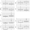
Box and whisker plots showing the distribution of parameters for control (Left) and PD (Right) populations for parameters determined to be significantly different.
FIGURE 3
FIGURE 3
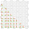
Lattice of cross-plots of statistically significant parameters colored by PD status (Green = Control). The upper diagonal shows correlation coefficients.
FIGURE 4
FIGURE 4
(A,B) Scanning electron microscopy of whole blood smears showing representative platelets from healthy individuals. (C–H) Whole blood smears from PD individuals showing hyperactivated platelets. (C–H) PD platelets agglutinating to RBCs; (D,H) PD platelet spreading (G) and PD platelet aggregation (C,E,F).
FIGURE 5
FIGURE 5
(A–G) Confocal microscopy images of PPP clots stained with the RgpA polyclonal antibody (1:100) from healthy individuals and individuals suffering from PD. The images (A–G) are two channel overlays where transmitted light microscopy micrograph and fluorescent signal are superimposed to show the areas of fluorescence on the clot itself, except (E) and (G) that show only the fluorescence signal. (A) The unstained and (B) stained control exhibits no fluorescent signal as well as (C) the unstained Parkinson’s disease PPP clots. (D–F) Fluorescent signal of the RgpA antibody is prominently detected in stained Parkinson’s disease PPP clots. (G) Represents a positive control in which a control sample that is absent of fluorescent signal received an exogenous load of RgpA. (H–L) Confocal microscopy images of fibrin networks formed from purified fibrinogen (with added Alexa488 fluorophore) incubated with and without RgpA, and LPS from P. gingivalis, followed by addition of thrombin to create extensive fibrin(ogen) clots. (H) Representative purified fibrin(ogen) clot. (I) A representative clot formed after purified fibrinogen was incubated with 10 ng L –1 P. gingivalis LPS. (J) A representative clot formed after purified fibrinogen was incubated with 100 ng L –1 RgpA and (K) 500 ng L –1 RgpA. (L) A representative clot after purified fibrinogen was simultaneously exposed to a combination of P. gingivalis LPS (10 ng L –1) and RgpA (500 ng L –1).
FIGURE 6
FIGURE 6
Box and whisker plot showing the distribution of mean RgpA image channel intensity for the healthy and PD populations.
FIGURE 7
FIGURE 7
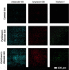
Examples of clots created with platelet poor plasma (PPP) for a representative control and two representative PD individuals to show amyloid fibrin(ogen) protein structure. Three fluorescent markers that bind amyloid protein were used, Amytracker 480, 680, and ThT (as previously used for amyloid fibrin structure (Pretorius et al., 2017c; de Waal et al., 2018).
FIGURE 8
FIGURE 8
Simplified platelet signaling and receptor activation with main dysregulated molecules IL-1α, IL-1 β, TNF-α, and IL-17A. When inflammatory molecules are upregulated in circulation, they either cause direct endothelial damage (by binding to receptors on endothelial cells), or they may act as ligands that bind directly to platelet membrane receptors (Olumuyiwa-Akeredolu et al., 2019). When these inflammatory molecules disrupt endothelial cell structure, the endothelial cells release collagen and von Willebrand Factor (vWF). vWF is also a mediator of vascular inflammation (Gragnano et al., 2017), and it binds to exposed collagen and anchors platelets to the subendothelium (Du, 2007), causing platelet aggregation (Xu et al., 2016), and formation of a platelet plug (Jagadapillai et al., 2016). Both collagen and vWF act as platelet receptor ligands, causing platelet outside-in signaling, followed by inside-out signaling. Furthermore, collagen and vWB binding also result in signaling processes that cause a release of stored molecules that are present inside α- and dense granules of platelets, and may also include stored interleukins (e.g., IL-6 and IL-1β); further increasing the concentration of these inflammatory molecules in circulation (Olumuyiwa-Akeredolu et al., 2019). vWF binding is mediated by GpIbα (which is part of the GPIb-IX-V) and integrin (αIIbβ3 complex (Bryckaert et al., 2015). This αIIbβ3 receptor also binds fibrinogen and thrombin, and both these molecules and vWF work together to play critical roles in platelet activation and aggregation (Estevez and Du, 2017). Diagram created using BioRender (https://biorender.com/).

References

    1. Aarabi G., Eberhard J., Reissmann D. R., Heydecke G., Seedorf U. (2015). Interaction between periodontal disease and atherosclerotic vascular disease–fact or fiction? Atherosclerosis 241 555–560. 10.1016/j.atherosclerosis.2015.04.819 - DOI - PubMed
    1. Ally N., Whisstock J. C., Sieprawska-Lupa M., Potempa J., Le Bonniec B. F., Travis J., et al. (2003). Characterization of the specificity of arginine-specific gingipains from Porphyromonas gingivalis reveals active site differences between different forms of the enzymes. Biochemistry 42 11693–11700. 10.1021/bi0349726 - DOI - PubMed
    1. Ambrosio N., Marin M. J., Laguna E., Herrera D., Sanz M., Figuero E., et al. (2019). Detection and quantification of Porphyromonas gingivalis and Aggregatibacter actinomycetemcomitans in bacteremia induced by interdental brushing in periodontally healthy and periodontitis patients. Arch. Oral Biol. 98 213–219. 10.1016/j.archoralbio.2018.11.025 - DOI - PubMed
    1. Anselmo A., Riva F., Gentile S., Soldani C., Barbagallo M., Mazzon C., et al. (2016). Expression and function of IL-1R8 (TIR8/SIGIRR): a regulatory member of the IL-1 receptor family in platelets. Cardiovasc. Res. 111 373–384. 10.1093/cvr/cvw162 - DOI - PubMed
    1. Ascherio A., Schwarzschild M. A. (2016). The epidemiology of parkinson’s disease: risk factors and prevention. Lancet Neurol. 15 1257–1272. 10.1016/s1474-4422(16)30230-7 - DOI - PubMed

LinkOut - more resources