

LPS Induces Preeclampsia-Like Phenotype in Rats and HTR8/SVneo Cells Dysfunction Through TLR4/p38 MAPK Pathway
- PMID: 31507429
- PMCID: PMC6718930
- DOI: 10.3389/fphys.2019.01030
LPS Induces Preeclampsia-Like Phenotype in Rats and HTR8/SVneo Cells Dysfunction Through TLR4/p38 MAPK Pathway
Abstract
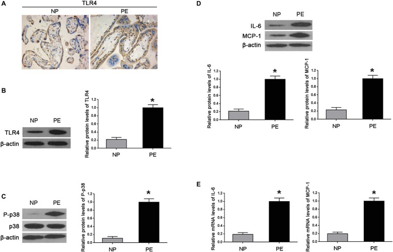
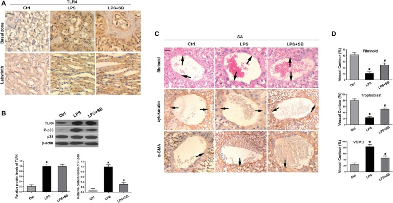
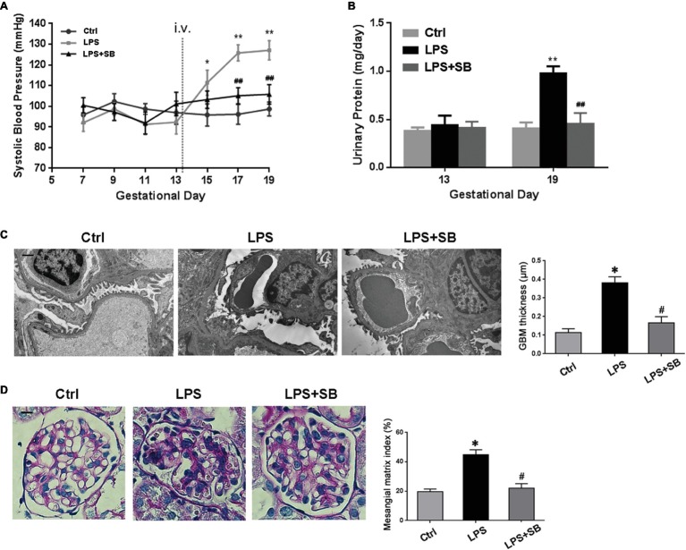
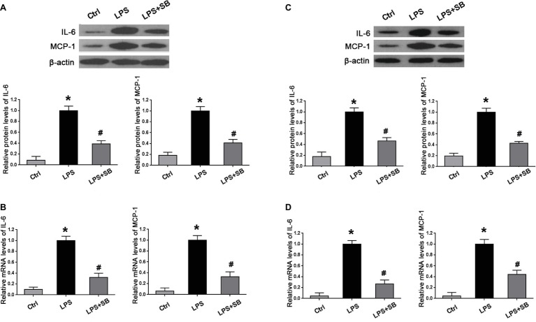
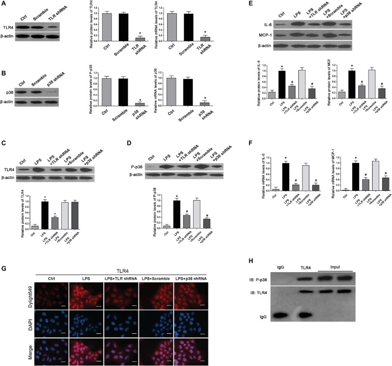
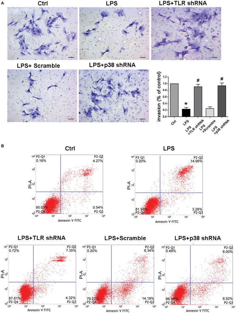
Accumulating evidence has shown that preeclampsia (PE) was associated with an aberrant maternal-fetal inflammatory response. In the present study, we first found that in human PE placentas levels of toll-like receptor 4 (TLR4), phosphorylated p38 mitogen-activated protein kinase (p-p38 MAPK) and inflammatory cytokines IL-6 and MCP-1 were significantly upregulated. Next, we demonstrated a notable increase in systolic blood pressure (SBP) and proteinuria in lipopolysaccharide (LPS)-treated pregnant rats and concomitant high levels of TLR4 and p-p38 in these PE-like rat placentas, which led to aberrant overexpression of both IL-6 and MCP-1, as well as deficient trophoblast invasion and spiral artery (SA) remodeling, and these abnormalities were ameliorated by SB203580, a reported inhibitor of p38. In vitro we further confirmed that LPS triggered the activation of TLR4/p38 signaling pathway, which promoted trophoblast apoptosis and damaged trophoblastic invasion via downstream effectors IL-6 and MCP-1; these mutations were rectified by silencing this signaling pathway. These findings elaborated potential mechanisms that aberrant TLR4/p38 signaling might contribute to PE and LPS-induced PE-like symptom by damaging trophoblast invasion and SA remodeling via activating inflammatory cytokines including IL-6 and MCP-1.
Keywords: inflammation; lipopolysaccharide; p38 MAPK; preeclampsia; spiral artery; toll-like receptor 4.
Figures
Similar articles
-
PAD4 silencing inhibits inflammation whilst promoting trophoblast cell invasion and migration by inactivating the NEMO/NF-κB pathway.Exp Ther Med. 2022 Jul 12;24(3):568. doi: 10.3892/etm.2022.11505. eCollection 2022 Sep. Exp Ther Med. 2022. PMID: 35978928 Free PMC article.
-
Curcumin improves LPS-induced preeclampsia-like phenotype in rat by inhibiting the TLR4 signaling pathway.Placenta. 2016 May;41:45-52. doi: 10.1016/j.placenta.2016.03.002. Epub 2016 Mar 10. Placenta. 2016. PMID: 27208407
-
Sevoflurane inhibited inflammatory response induced by TNF-α in human trophoblastic cells through p38MAPK signaling pathway.J Recept Signal Transduct Res. 2020 Jun;40(3):218-223. doi: 10.1080/10799893.2020.1726951. Epub 2020 Feb 18. J Recept Signal Transduct Res. 2020. PMID: 32069432
-
Single administration of ultra-low-dose lipopolysaccharide in rat early pregnancy induces TLR4 activation in the placenta contributing to preeclampsia.PLoS One. 2015 Apr 8;10(4):e0124001. doi: 10.1371/journal.pone.0124001. eCollection 2015. PLoS One. 2015. PMID: 25853857 Free PMC article.
-
Oxidative stress-induced Gadd45α inhibits trophoblast invasion and increases sFlt1/sEng secretions via p38 MAPK involving in the pathology of pre-eclampsia.J Matern Fetal Neonatal Med. 2016 Dec;29(23):3776-85. doi: 10.3109/14767058.2016.1144744. Epub 2016 Mar 3. J Matern Fetal Neonatal Med. 2016. PMID: 26809169
Cited by
-
Advanced platelet-rich fibrin promotes the paracrine function and proliferation of adipose-derived stem cells and contributes to micro-autologous fat transplantation by modulating HIF-1α and VEGF.Ann Transl Med. 2022 Jan;10(2):60. doi: 10.21037/atm-21-6812. Ann Transl Med. 2022. PMID: 35282074 Free PMC article.
-
Inflammation-mediated generation and inflammatory potential of human placental cell-free fetal DNA.Placenta. 2020 Apr;93:49-55. doi: 10.1016/j.placenta.2020.02.016. Epub 2020 Feb 24. Placenta. 2020. PMID: 32250739 Free PMC article.
-
LncRNA NEAT1 Promotes TLR4 Expression to Regulate Lipopolysaccharide-Induced Trophoblastic Cell Pyroptosis as a Molecular Sponge of miR-302b-3p.Mol Biotechnol. 2022 Jun;64(6):670-680. doi: 10.1007/s12033-021-00436-2. Epub 2022 Jan 22. Mol Biotechnol. 2022. PMID: 35064469
-
Establishment and comparison of human term placenta-derived trophoblast cells†.Biol Reprod. 2024 May 9;110(5):950-970. doi: 10.1093/biolre/ioae026. Biol Reprod. 2024. PMID: 38330185 Free PMC article.
-
PAD4 silencing inhibits inflammation whilst promoting trophoblast cell invasion and migration by inactivating the NEMO/NF-κB pathway.Exp Ther Med. 2022 Jul 12;24(3):568. doi: 10.3892/etm.2022.11505. eCollection 2022 Sep. Exp Ther Med. 2022. PMID: 35978928 Free PMC article.
References
-
- Bernardi F. C., Felisberto F., Vuolo F., Petronilho F., Souza D. R., Luciano T. F., et al. . (2012). Oxidative damage, inflammation, and Toll-like receptor 4 pathway are increased in preeclamptic patients: a case-control study. Oxidative Med. Cell. Longev. 2012:636419. 10.1155/2012/636419, PMID: - DOI - PMC - PubMed
-
- Brosens I. A., Robertson W. B., Dixon H. G. (1972). The role of the spiral arteries in the pathogenesis of preeclampsia. Obstet. Gynecol. Annu. 1, 177–191. - PubMed
LinkOut - more resources
Full Text Sources
Miscellaneous