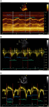
Comparison of Different Methods for Estimating Cardiac Timings: A Comprehensive Multimodal Echocardiography Investigation
- PMID: 31507437
- PMCID: PMC6713915
- DOI: 10.3389/fphys.2019.01057
Comparison of Different Methods for Estimating Cardiac Timings: A Comprehensive Multimodal Echocardiography Investigation
Abstract
Cardiac time intervals are important hemodynamic indices and provide information about left ventricular performance. Phonocardiography (PCG), impedance cardiography (ICG), and recently, seismocardiography (SCG) have been unobtrusive methods of choice for detection of cardiac time intervals and have potentials to be integrated into wearable devices. The main purpose of this study was to investigate the accuracy and precision of beat-to-beat extraction of cardiac timings from the PCG, ICG and SCG recordings in comparison to multimodal echocardiography (Doppler, TDI, and M-mode) as the gold clinical standard. Recordings were obtained from 86 healthy adults and in total 2,120 cardiac cycles were analyzed. For estimation of the pre-ejection period (PEP), 43% of ICG annotations fell in the corresponding echocardiography ranges while this was 86% for SCG. For estimation of the total systolic time (TST), these numbers were 43, 80, and 90% for ICG, PCG, and SCG, respectively. In summary, SCG and PCG signals provided an acceptable accuracy and precision in estimating cardiac timings, as compared to ICG.
Keywords: cardiac time intervals; echocardiography; impedance cardiography (ICG); left ventricular ejection time (LVET); phonocardiography (PCG); pre-ejection period (PEP); seismocardiography (SCG).
Figures


Similar articles
-
Estimation of Beat-to-Beat Interval and Systolic Time Intervals Using Phono- and Seismocardiograms.Annu Int Conf IEEE Eng Med Biol Soc. 2019 Jul;2019:5650-5656. doi: 10.1109/EMBC.2019.8856931. Annu Int Conf IEEE Eng Med Biol Soc. 2019. PMID: 31947135
-
Wearable seismocardiography: towards a beat-by-beat assessment of cardiac mechanics in ambulant subjects.Auton Neurosci. 2013 Nov;178(1-2):50-9. doi: 10.1016/j.autneu.2013.04.005. Epub 2013 May 9. Auton Neurosci. 2013. PMID: 23664242
-
Comparison of the measured pre-ejection periods and left ventricular ejection times between echocardiography and impedance cardiography for optimizing cardiac resynchronization therapy.J Arrhythm. 2017 Apr;33(2):130-133. doi: 10.1016/j.joa.2016.08.003. Epub 2016 Sep 12. J Arrhythm. 2017. PMID: 28416980 Free PMC article.
-
Holter-type impedance cardiography device. A system for continuous and non-invasive monitoring of cardiac haemodynamics.Kardiol Pol. 2004 Aug;61(8):138-46. Kardiol Pol. 2004. PMID: 15457280 Review.
-
Systolic Time Intervals and New Measurement Methods.Cardiovasc Eng Technol. 2016 Jun;7(2):118-25. doi: 10.1007/s13239-016-0262-1. Epub 2016 Apr 5. Cardiovasc Eng Technol. 2016. PMID: 27048269 Review.
Cited by
-
Cardiac function and posttraumatic stress disorder: a review of the literature and case report.Health Promot Chronic Dis Prev Can. 2023 Nov;43(10-11):472-480. doi: 10.24095/hpcdp.43.10/11.05. Health Promot Chronic Dis Prev Can. 2023. PMID: 37991890 Free PMC article. Review.
-
Novel Approach to Assess Cardiac Function Using Systolic Performance and Myocardial Performance Indices From Simultaneous Electrocardiography and Phonocardiography Recordings in Dogs With Various Stages of Myxomatous Mitral Valve Disease.Front Vet Sci. 2021 Oct 21;8:741115. doi: 10.3389/fvets.2021.741115. eCollection 2021. Front Vet Sci. 2021. PMID: 34746282 Free PMC article.
-
Validity and reliability of seismocardiography for the estimation of cardiorespiratory fitness.Cardiovasc Digit Health J. 2023 Sep 11;4(5):155-163. doi: 10.1016/j.cvdhj.2023.08.020. eCollection 2023 Oct. Cardiovasc Digit Health J. 2023. PMID: 37850043 Free PMC article.
-
A Wearable Multi-Sensor Array Enables the Recording of Heart Sounds in Homecare.Sensors (Basel). 2023 Jul 7;23(13):6241. doi: 10.3390/s23136241. Sensors (Basel). 2023. PMID: 37448089 Free PMC article.
-
Detecting Coronary Artery Disease Using Rest Seismocardiography and Gyrocardiography.Front Physiol. 2021 Dec 2;12:758727. doi: 10.3389/fphys.2021.758727. eCollection 2021. Front Physiol. 2021. PMID: 34925059 Free PMC article.
References
-
- Baevskii R. M., Egorov A. D., Kazarian L. A. (1964). The method of seismocardiography. Kardiologiia 18, 87–89. - PubMed
LinkOut - more resources
Full Text Sources
Other Literature Sources
Medical
Miscellaneous

