

Networks Disrupted in Linguistic Variants of Frontotemporal Dementia
- PMID: 31507513
- PMCID: PMC6716200
- DOI: 10.3389/fneur.2019.00903
Networks Disrupted in Linguistic Variants of Frontotemporal Dementia
Abstract
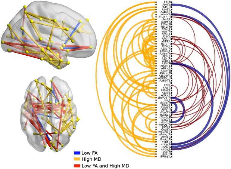
The non-fluent/agrammatic variant of primary progressive aphasia (nfvPPA) and semantic variant (svPPA) of frontotemporal dementia (FTD) are neurodegenerative diseases. Previous works have shown alterations of fractional anisotropy (FA) and mean diffusivity (MD) from diffusion tensor images (DTIs). This manuscript is aimed at using DTI images to build a global tractography and to identify atrophy patterns of white matter in each variant. Twenty patients with svPPA, 20 patients with nfvPPA, 26 patients with behavioral variant of FTD (bvFTD) and, 33 controls were included. An analysis based on the connectivity of structural networks showed changes in FA and MD in svPPA and nfvPPA with respect to bvFTD. Much damage in the internal networks of the left temporal lobe was found in svPPA patients; in contrast, patients with nfvPPA showed atrophy in networks from the basal ganglia to motor and premotor areas. Those findings support the dual stream model of speech and language.
Keywords: diffusion tensor imaging; fractional anisotropy; frontotemporal dementia; primary progressive aphasia; structural connectivity; white matter.
Figures


Similar articles
-
Longitudinal white matter change in frontotemporal dementia subtypes and sporadic late onset Alzheimer's disease.Neuroimage Clin. 2017 Sep 14;16:595-603. doi: 10.1016/j.nicl.2017.09.007. eCollection 2017. Neuroimage Clin. 2017. PMID: 28975068 Free PMC article.
-
Modelling pathological spread through the structural connectome in the frontotemporal dementia clinical spectrum.Brain. 2025 Jun 3;148(6):1994-2007. doi: 10.1093/brain/awae391. Brain. 2025. PMID: 39611765 Free PMC article.
-
Infratentorial pathology in frontotemporal dementia: cerebellar grey and white matter alterations in FTD phenotypes.J Neurol. 2021 Dec;268(12):4687-4697. doi: 10.1007/s00415-021-10575-w. Epub 2021 May 13. J Neurol. 2021. PMID: 33983551 Free PMC article.
-
Frontotemporal lobar degeneration: a clinical approach.Semin Neurol. 2014 Apr;34(2):189-201. doi: 10.1055/s-0034-1381735. Epub 2014 Jun 25. Semin Neurol. 2014. PMID: 24963678 Review.
-
The Role of Graph Theory in Evaluating Brain Network Alterations in Frontotemporal Dementia.Front Neurol. 2022 Jun 28;13:910054. doi: 10.3389/fneur.2022.910054. eCollection 2022. Front Neurol. 2022. PMID: 35837233 Free PMC article. Review.
Cited by
-
Assessing processing speed and its neural correlates in the three variants of primary progressive aphasia with a non-verbal tablet-based task.Cortex. 2024 Feb;171:165-177. doi: 10.1016/j.cortex.2023.10.011. Epub 2023 Nov 2. Cortex. 2024. PMID: 38000139 Free PMC article.
-
Speech and language impairments in behavioral variant frontotemporal dementia: A systematic review.Neurosci Biobehav Rev. 2021 Dec;131:1076-1095. doi: 10.1016/j.neubiorev.2021.10.015. Epub 2021 Oct 19. Neurosci Biobehav Rev. 2021. PMID: 34673112 Free PMC article.
-
Altered structural brain networks in linguistic variants of frontotemporal dementia.Brain Imaging Behav. 2022 Jun;16(3):1113-1122. doi: 10.1007/s11682-021-00560-2. Epub 2021 Nov 10. Brain Imaging Behav. 2022. PMID: 34755293 Free PMC article.
-
Multiparametric MRI-based biomarkers in the non-fluent and semantic variants of primary progressive aphasia.J Neurol. 2025 Jul 2;272(8):490. doi: 10.1007/s00415-025-13215-9. J Neurol. 2025. PMID: 40601042 Free PMC article.
-
Asymmetry of radiomics features in the white matter of patients with primary progressive aphasia.Front Aging Neurosci. 2023 May 5;15:1120935. doi: 10.3389/fnagi.2023.1120935. eCollection 2023. Front Aging Neurosci. 2023. PMID: 37213534 Free PMC article.
References
LinkOut - more resources
Full Text Sources