. 2019 Aug 22:10:911.
doi: 10.3389/fneur.2019.00911. eCollection 2019.

Light-Emitting Diode Photobiomodulation After Cerebral Ischemia

Affiliations

Light-Emitting Diode Photobiomodulation After Cerebral Ischemia

Bárbara Argibay et al. Front Neurol. .

Abstract

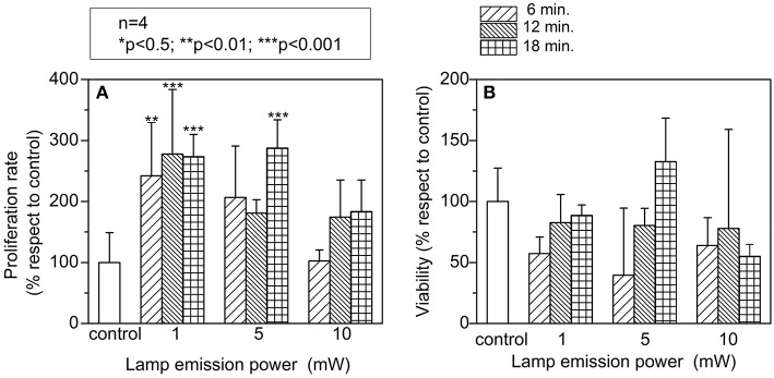
Photobiomodulation (PBM) therapy is a promising therapeutic approach for several pathologies, including stroke. The biological effects of PBM for the treatment of cerebral ischemia have previously been explored as a neuroprotective strategy using different light sources, wavelengths, and incident light powers. However, the capability of PBM as a novel alternative therapy to stimulate the recovery of the injured neuronal tissue after ischemic stroke has been poorly explored. The aim of this study was to investigate the low-level light irradiation therapy by using Light Emitting Diodes (LEDs) as potential therapeutic strategy for stroke. The LED photobiomodulation (continuous wave, 830 nm, 0.2-0.6 J/cm2) was firstly evaluated at different energy densities in C17.2 immortalized mouse neural progenitor cell lines, in order to observe if this treatment had any effect on cells, in terms of proliferation and viability. Then, the PBM-LED effect (continuous wave, 830 nm, 0.28 J/cm2 at brain cortex) on long-term recovery (12 weeks) was analyzed in ischemic animal model by means lesion reduction, behavioral deficits, and functional magnetic resonance imaging (fMRI). Analysis of cellular proliferation after PBM was significantly increased (1 mW) in all different exposure times used; however, this effect could not be replicated in vivo experimental conditions, as PBM did not show an infarct reduction or functional recovery. Despite the promising therapeutic effect described for PBM, further preclinical studies are necessary to optimize the therapeutic window of this novel therapy, in terms of the mechanism associated to neurorecovery and to reduce the risk of failure in futures clinical trials.

Keywords: animal model; functional recovery; intracerebral hemorrhage; ischemic stroke; magnetic resonance imaging; photobiomodulation therapy.


Figures

Figure 1
Figure 1
In vitro study to evaluate: (A) cellular proliferation; cell count was found significant increased at low emission powers and exposures times. (B) Viability; photostimulaltion did not produce toxic side effects in vitro. A two-way analysis of variance (ANOVA) followed by post-hoc Bonferroni evaluation was used. (n = 4; *p < 0.5; **p < 0.01; ***p < 0.001). Lamp emission power at three different exposure time (6, 12, and 18 min).
Figure 2
Figure 2
Functional brain recovery observation. (A) Representative BOLD images obtained during pre-cerebral ischemia, 5 and 12 weeks after the insult in both hemispheres. (B) Alternating unilateral forepaw stimulation was performed using rectangular pulses. BOLD fMRI was conducted alternating three times between each hemisphere, and animals were allowed to rest for 10 min between stimulation sessions. The time course of each pixel during forepaw stimulation was examined using a paired Student's t-test (p < 0.05). Only clusters that included at least four adjacent activated pixels were considered as positive activation areas. (n = 6 animals/group). All animals exhibited BOLD signal in both hemispheres (measured separately) prior to the cerebral ischemia. The animals were followed for 12 weeks and all of them exhibited BOLD signal in the contralateral side but none of them showed BOLD activation in the ipsilateral side.
Figure 3
Figure 3
In vivo study: (A) Cylinder test results obtained from the three studied groups. (B) Ischemic volume results of the three groups; No significant differences were observed between groups up to 12 weeks. (C) T2-weighted MR images of a representative brain of each photostimulated group.
Figure 4
Figure 4
Histological analysis of neurons (Fox3), astrocytes (GFAP), and neurogenesis (DCX and Ki 67) of animals from each experimental group at 12 weeks after ischemic lesion. Cortical (*) and subcortical brain regions (**).

References

    1. Oron A, Oron U, Chen J, Eilam A, Zhang C, Sadeh M, et al. . Low-level laser therapy applied transcranially to rats after induction of stroke significantly reduces long-term neurological deficits. Stroke. (2006) 37:2620–4. 10.1161/01.STR.0000242775.14642.b8 - DOI - PubMed
    1. Hu WP, Wang JJ, Yu CL, Lan CC, Chen GS, Yu HS. Helium-neon laser irradiation stimulates cell proliferation through photostimulatory effects in mitochondria. J Invest Dermatol. (2007) 127:2048–57. 10.1038/sj.jid.5700826 - DOI - PubMed
    1. Huang YY, Sharma SK, Carroll J, Hamblin MR. Biphasic dose response in low level light therapy - an update. Dose Response. (2011) 9:602–18. 10.2203/dose-response.11-009.Hamblin - DOI - PMC - PubMed
    1. Thunshelle C, Hamblin MR. Transcranial low-level laser (light) therapy for brain injury. Photomed Laser Surg. (2016) 34:587–98. 10.1089/pho.2015.4051 - DOI - PMC - PubMed
    1. Salehpour F, Mahmoudi J, Kamari F, Sadigh-Eteghad S, Rasta SH, Hamblin MR. Brain photobiomodulation therapy: a narrative review. Mol Neurobiol. (2018) 55:6601–36. 10.1007/s12035-017-0852-4 - DOI - PMC - PubMed

LinkOut - more resources