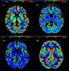
Multimodal Quantitative MRI Reveals No Evidence for Tissue Pathology in Idiopathic Cervical Dystonia
- PMID: 31507518
- PMCID: PMC6719627
- DOI: 10.3389/fneur.2019.00914
Multimodal Quantitative MRI Reveals No Evidence for Tissue Pathology in Idiopathic Cervical Dystonia
Abstract
Background: While in symptomatic forms of dystonia cerebral pathology is by definition present, it is unclear so far whether disease is associated with microstructural cerebral changes in idiopathic dystonia. Previous quantitative MRI (qMRI) studies assessing cerebral tissue composition in idiopathic dystonia revealed conflicting results. Objective: Using multimodal qMRI, the presented study aimed to investigate alterations in different cerebral microstructural compartments associated with idiopathic cervical dystonia in vivo. Methods: Mapping of T1, T2, , and proton density (PD) was performed in 17 patients with idiopathic cervical dystonia and 29 matched healthy control subjects. Statistical comparisons of the parametric maps between groups were conducted for various regions of interest (ROI), including major basal ganglia nuclei, the thalamus, white matter, and the cerebellum, and voxel-wise for the whole brain. Results: Neither whole brain voxel-wise statistics nor ROI-based analyses revealed significant group differences for any qMRI parameter under investigation. Conclusions: The negative findings of this qMRI study argue against the presence of overt microstructural tissue change in patients with idiopathic cervical dystonia. The results seem to support a common view that idiopathic cervical dystonia might primarily resemble a functional network disease.
Keywords: idiopathic dystonia; movement disorders; proton density; quantitative MRI; relaxometry.
Figures


Similar articles
-
Focal epilepsy without overt epileptogenic lesions: no evidence of microstructural brain tissue damage in multi-parametric quantitative MRI.Front Neurol. 2023 Jul 17;14:1175971. doi: 10.3389/fneur.2023.1175971. eCollection 2023. Front Neurol. 2023. PMID: 37528856 Free PMC article.
-
A group-level comparison of volumetric and combined volumetric-surface normalization for whole brain analyses of myelin and iron maps.Magn Reson Imaging. 2018 Dec;54:225-240. doi: 10.1016/j.mri.2018.08.021. Epub 2018 Sep 1. Magn Reson Imaging. 2018. PMID: 30176374
-
Basal ganglia and cerebellar pathology in X-linked dystonia-parkinsonism.Brain. 2018 Oct 1;141(10):2995-3008. doi: 10.1093/brain/awy222. Brain. 2018. PMID: 30169601
-
Secondary cervical dystonia associated with structural lesions of the central nervous system.Mov Disord. 2003 Jan;18(1):60-9. doi: 10.1002/mds.10301. Mov Disord. 2003. PMID: 12518301 Review.
-
Pathology of idiopathic dystonia: findings from genetic animal models.Prog Neurobiol. 1998 Apr;54(6):633-77. doi: 10.1016/s0301-0082(97)00089-0. Prog Neurobiol. 1998. PMID: 9560845 Review.
Cited by
-
Gray matter structural alterations of cortico-striato-thalamo-cortical loop in familial Paroxysmal Kinesigenic Dyskinesia.Heliyon. 2024 Aug 22;10(17):e36739. doi: 10.1016/j.heliyon.2024.e36739. eCollection 2024 Sep 15. Heliyon. 2024. PMID: 39263125 Free PMC article.
-
The Central Effects of Botulinum Toxin in Dystonia and Spasticity.Toxins (Basel). 2021 Feb 17;13(2):155. doi: 10.3390/toxins13020155. Toxins (Basel). 2021. PMID: 33671128 Free PMC article. Review.
-
No reliable gray matter alterations in idiopathic dystonia.Front Neurol. 2025 Mar 3;16:1510115. doi: 10.3389/fneur.2025.1510115. eCollection 2025. Front Neurol. 2025. PMID: 40098684 Free PMC article.
-
Voxel-based meta-analysis of gray matter abnormalities in idiopathic dystonia.J Neurol. 2022 Jun;269(6):2862-2873. doi: 10.1007/s00415-022-10961-y. Epub 2022 Jan 11. J Neurol. 2022. PMID: 35013788
-
Multiparametric Quantitative MRI in Neurological Diseases.Front Neurol. 2021 Mar 8;12:640239. doi: 10.3389/fneur.2021.640239. eCollection 2021. Front Neurol. 2021. PMID: 33763021 Free PMC article. Review.
References
LinkOut - more resources
Full Text Sources

