Parcellation of the Hippocampus Using Resting Functional Connectivity in Temporal Lobe Epilepsy
- PMID: 31507522
- PMCID: PMC6714062
- DOI: 10.3389/fneur.2019.00920
Parcellation of the Hippocampus Using Resting Functional Connectivity in Temporal Lobe Epilepsy
Abstract
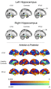
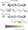
We have previously shown that the connectivity of the hippocampus to other regions of the default mode network (DMN) is a strong indicator of memory ability in people with temporal lobe epilepsy (TLE). Recent work in the cognitive neuroscience literature has suggested that the anterior and posterior aspects of the hippocampus have distinct connections to the rest of the DMN and may support different memory operations. Further, structural analysis of epileptogenic hippocampi has found greater atrophy, characterized by mesial temporal sclerosis, in the anterior region of the hippocampus. Here, we used resting state FMRI data to parcellate the hippocampus according to its functional connectivity to the rest of the brain in people with left lateralized TLE (LTLE) and right lateralized TLE (RTLE), and in a group of neurologically healthy controls. We found similar anterior and posterior compartments in all groups. However, there was weaker connectivity of the epileptogenic hippocampus to multiple regions of the DMN. Both TLE groups showed reduced connectivity of the posterior hippocampus to key hubs of the DMN, the posterior cingulate cortex (PCC) and the medial pre-frontal cortex (mPFC). In the LTLE group, the anterior hippocampus also showed reduced connectivity to the DMN, and this effect was influenced by the presence of mesial temporal sclerosis. When we explored brain-behavior relationships, we found that reduced connectivity of the left anterior hippocampus to the DMN hubs related to poorer verbal memory ability in people with LTLE, and reduced connectivity of the right posterior hippocampus to the PCC related to poorer visual memory ability in those with RTLE. These findings may inform models regarding functional distinctions of the hippocampal anteroposterior axis.
Keywords: default mode network; epilepsy; hippocampus; long axis; memory; resting state.
Figures


Similar articles
-
Diminished default mode network recruitment of the hippocampus and parahippocampus in temporal lobe epilepsy.J Neurosurg. 2013 Aug;119(2):288-300. doi: 10.3171/2013.3.JNS121041. Epub 2013 May 24. J Neurosurg. 2013. PMID: 23706058 Free PMC article.
-
Resting-state hippocampal networks related to language processing reveal unique patterns in temporal lobe epilepsy.Epilepsy Behav. 2021 Apr;117:107834. doi: 10.1016/j.yebeh.2021.107834. Epub 2021 Feb 18. Epilepsy Behav. 2021. PMID: 33610102 Free PMC article.
-
Patterns of default mode network in temporal lobe epilepsy with and without hippocampal sclerosis.Epilepsy Behav. 2021 Aug;121(Pt B):106523. doi: 10.1016/j.yebeh.2019.106523. Epub 2019 Oct 20. Epilepsy Behav. 2021. PMID: 31645315
-
The structural and functional connectivity of the posterior cingulate cortex: comparison between deterministic and probabilistic tractography for the investigation of structure-function relationships.Neuroimage. 2014 Nov 15;102 Pt 1:118-27. doi: 10.1016/j.neuroimage.2013.12.022. Epub 2013 Dec 21. Neuroimage. 2014. PMID: 24365673 Review.
-
Mechanisms of cognitive impairment in temporal lobe epilepsy: A systematic review of resting-state functional connectivity studies.Epilepsy Behav. 2021 Feb;115:107686. doi: 10.1016/j.yebeh.2020.107686. Epub 2020 Dec 24. Epilepsy Behav. 2021. PMID: 33360743
Cited by
-
Schemas provide a scaffold for neocortical integration of new memories over time.Nat Commun. 2022 Oct 2;13(1):5795. doi: 10.1038/s41467-022-33517-0. Nat Commun. 2022. PMID: 36184668 Free PMC article.
-
Dynamic functional connectivity in modular organization of the hippocampal network marks memory phenotypes in temporal lobe epilepsy.Hum Brain Mapp. 2022 Apr 15;43(6):1917-1929. doi: 10.1002/hbm.25763. Epub 2021 Dec 30. Hum Brain Mapp. 2022. PMID: 34967488 Free PMC article.
-
Hippocampal anterior- posterior shift in childhood and adolescence.Prog Neurobiol. 2023 Jun;225:102447. doi: 10.1016/j.pneurobio.2023.102447. Epub 2023 Mar 24. Prog Neurobiol. 2023. PMID: 36967075 Free PMC article.
-
Data-Driven Clustering of Functional Signals Reveals Gradients in Processing Both within the Anterior Hippocampus and across Its Long Axis.J Neurosci. 2022 Sep 28;42(39):7431-7441. doi: 10.1523/JNEUROSCI.0269-22.2022. J Neurosci. 2022. PMID: 36002264 Free PMC article.
-
An exploratory study into the influence of laterality and location of hippocampal sclerosis on seizure prognosis and global cortical thinning.Sci Rep. 2021 Feb 25;11(1):4686. doi: 10.1038/s41598-021-84281-y. Sci Rep. 2021. PMID: 33633325 Free PMC article.
References
LinkOut - more resources
Full Text Sources

