. 2019 Aug 27:10:2006.
doi: 10.3389/fmicb.2019.02006. eCollection 2019.

Gga-miR-19b-3p Inhibits Newcastle Disease Virus Replication by Suppressing Inflammatory Response via Targeting RNF11 and ZMYND11

Affiliations

Gga-miR-19b-3p Inhibits Newcastle Disease Virus Replication by Suppressing Inflammatory Response via Targeting RNF11 and ZMYND11

Yu Chen et al. Front Microbiol. .

Abstract

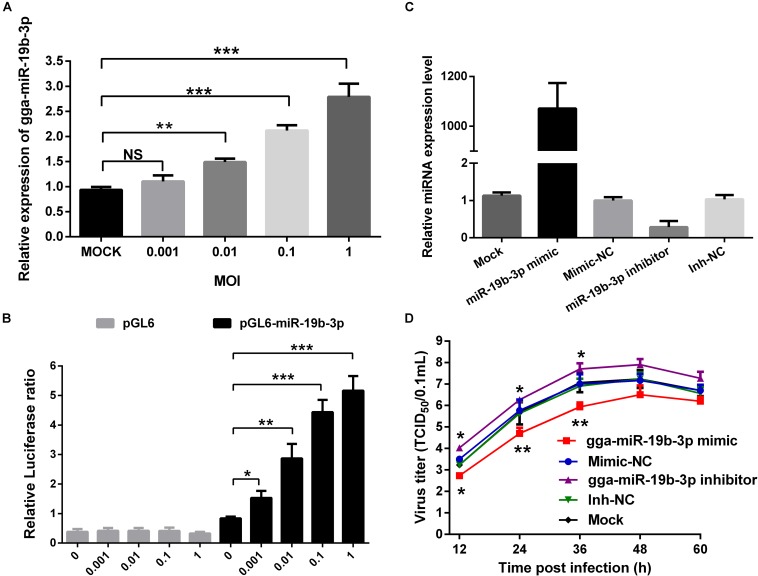
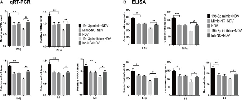
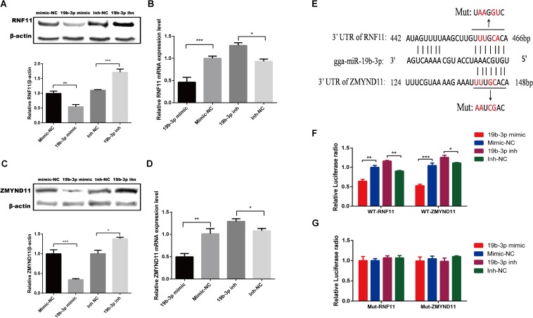
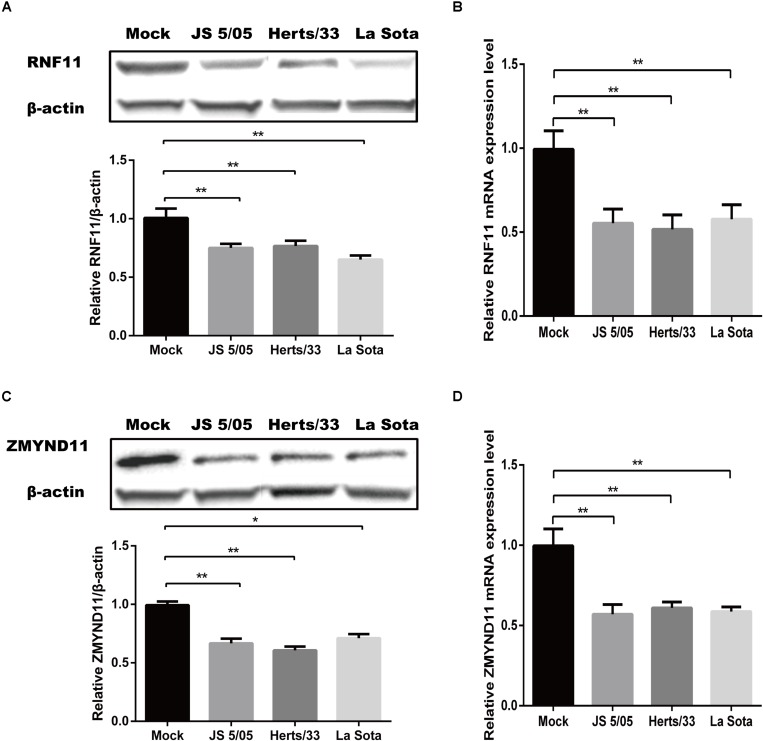
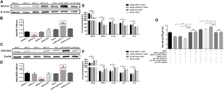
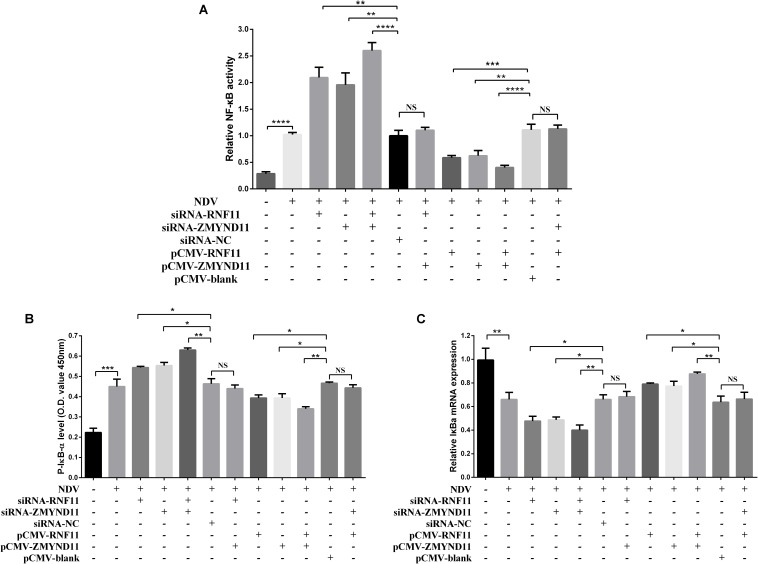
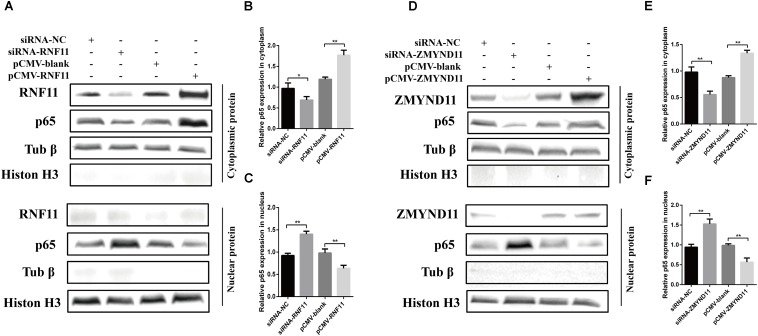
Newcastle disease (ND), an acute and highly contagious avian disease caused by virulent Newcastle disease virus (NDV), often results in severe economic losses worldwide every year. Although it is clear that microRNAs (miRNAs) are implicated in modulating innate immune response to invading microbial pathogens, their role in host defense against NDV infection remains largely unknown. Our prior study indicates that gga-miR-19b-3p is up-regulated in NDV-infected DF-1 cells (a chicken embryo fibroblast cell line) and functions to suppress NDV replication. Here we report that overexpression of gga-miR-19b-3p promoted the production of NDV-induced inflammatory cytokines and suppressed NDV replication, whereas inhibition of endogenous gga-miR-19b-3p expression had an opposite effect. Dual-luciferase and gene expression array analyses revealed that gga-miR-19b-3p directly targets the mRNAs of ring finger protein 11 (RNF11) and zinc-finger protein, MYND-type containing 11 (ZMYND11), two negative regulators of nuclear factor kappa B (NF-κB) signaling, in DF-1 cells. RNF11 and ZMYND11 silencing by small interfering RNA (siRNA) induced NF-κB activity and inflammatory cytokine production, and suppressed NDV replication; whereas ectopic expression of these two proteins exhibited an opposite effect. Our study provides evidence that gga-miR-19b-3p activates NF-κB signaling by targeting RNF11 and ZMYND11, and that enhanced inflammatory cytokine production is likely responsible for the suppression of NDV replication.

Keywords: DF-1 cells; NDV; NF-κB signaling; RNF11; ZMYND11; gga-miR-19b-3p; inflammatory cytokine.


Figures

FIGURE 1
FIGURE 1
Newcastle disease virus-induced gga-miR-19b-3p inhibits NDV replication in DF-1 cells. (A) DF-1 cells were infected with JS 5/05 strain at indicated MOIs for 12 h, and then the expression of gga-miR-19b-3p was detected by qRT-PCR. (B) The pGL-6-miR-19b-3p or pGL-6 plasmid was transfected into DF-1 cells together with pRL-TK renilla luciferase reporter plasmid as an internal control. Forty-eight hours post transfection, the cells were infected with JS 5/05 strain at different MOIs for 12 h, and then the dual-luciferase activity was detected and firefly luciferase activities were normalized to firefly renilla luciferase activities. (C,D) DF-1 cells were transfected with gga-miR-19b-3p mimic or inhibitor or mimic-NC or inh-NC at a final concentration of 100 nM or left untreated. The gga-miR-19b-3p expression was measured by qRT-PCR at 18 h post transfection (C). Eighteen hours after transfection, DF-1 cells were infected with JS5/05 strain at an MOI of 0.1. The viral road in the supernatants collected at different time points were quantified by TCID50 on DF-1 cells (D). Results are representative of three independent experiments and presented as means ± SD. p < 0.05, ∗∗p < 0.01, ∗∗∗p < 0.001.
FIGURE 2
FIGURE 2
Gga-miR-19b-3p promotes NDV-induced expression of inflammatory cytokines. DF-1 cells were transfected with indicated RNA oligonucleotides at a final concentration of 100 nM for 18 h or left untreated before infected with JS 5/05 at an MOI of 0.1. The expression levels of IFN-β, TNF-α, IL-1β, IL-6, and IL-8 were measured by qRT-PCR (A) and ELISA (B) at 18 hpi. Results are representative of three independent experiments and presented as means ± SD. p < 0.05, ∗∗p < 0.01, ∗∗∗p < 0.001.
FIGURE 3
FIGURE 3
Gga-miR-19b-3p directly targets RNF11 and ZMYND11 in DF-1 cells. DF-1 cells were transfected with indicated RNA oligonucleotides at a final concentration of 100 nM for 18 h. Then the expression of RNF11 and ZMYND11 were detected by Western blot (A,C) and qRT-PCR (B,D) assays. The relative levels of RNF11 and ZMYND11 proteins were calculated as follows: band density of RNF11 or ZMYND11/band density of β-actin in the same sample and showed in the below of (A,C). Diagram of the predicted target sites for gga-miR-19b-3p in RNF11 and ZMYND11 were shown in (E). DF-1 cells were co-transfected with indicated RNA oligonucleotides and wild type (F) or mutant luciferase reporter gene vectors (G) before luciferase reporter gene assay was performed. The relative level of renilla luciferase activities was calculated normalized on the basis of firefly luciferase activities. Data are representative of three independent experiments and presented as means ± SD. p < 0.05, ∗∗p < 0.01, ∗∗∗p < 0.001.
FIGURE 4
FIGURE 4
Newcastle disease virus infection inhibits the expressions of RNF11 and ZMYND11. DF-1 cells were infected with different NDV strains (JS 5/05, Herts/33, and La Sota) at an MOI of 0.1 for 18 h. The expressions of RNF11 and ZMYND11 were detected by Western blot (A,C) and qRT-PCR (B,D) assays. The relative levels of RNF11 and ZMYND11 proteins were calculated as described above and showed in the below of (A,C). Data are representative of three independent experiments and presented as means ± SD. p < 0.05, ∗∗p < 0.01.
FIGURE 5
FIGURE 5
RNF11 and ZMYND11 inhibit NDV-induced inflammatory cytokines and promote NDV replication in DF-1 cells. DF-1 cells were transfected with indicated siRNAs or siRNA-NC or overexpression plasmids or pCMV-blank. The efficiencies of interference and overexpression in transfected DF-1 cells were measured by Western blot (A,C) and qRT-PCR (B,D) at 18 h post transfection. DF-1 cells transfected with indicated siRNAs or siRNA-NC or overexpression plasmids or pCMV-blank or left untreated were infected with JS 5/05 at an MOI of 0.1 for 18 h. The expression levels of IFN-β, TNF-α, IL-1β, IL-6, and IL-8 were measured by ELISA assay (E,F). DF-1 cells transfected with indicated siRNAs or siRNA-NC or overexpression plasmids or pCMV-blank were infected with JS 5/05 at an MOI of 0.1. After 36 h, viral titers in infected DF-1 cells were measured by TCID50 as described above (G). Data are representative of three independent experiments and presented as means ± SD. p < 0.05, ∗∗p < 0.01, ∗∗∗p < 0.001.
FIGURE 6
FIGURE 6
RNF11 and ZMYND11 suppress NF-κB activity. pNF-κB-luc plasmid and indicated siRNAs or siRNA-NC or overexpression plasmids or pCMV-blank were co-transfected into DF-1 cells together with the internal control plasmid pRL-TK. Eighteen hours after transfection, the cells were infected with JS 5/05 strain at an MOI of 0.1 or left uninfected, dual-luciferase (A), ELISA (B), and qRT-PCR (C) assays were performed as described above at 18 hpi. NF-κB activities were indicated by the ratio of firefly luciferase activities to renilla luciferase activities. The relative expression of IκB-α was normalized with GAPDH and calculated using the 2–Δ Δ CT method. Data are representative of three independent experiments and presented as means ± SD. p < 0.05, ∗∗p < 0.01, ∗∗∗p < 0.001, *⁣*⁣**p < 0.0001.
FIGURE 7
FIGURE 7
RNF11 and ZMYND11 inhibit the nucleus translocation of p65. DF-1 cells were transfected with indicated siRNAs or siRNA-NC or overexpression plasmids or pCMV-blank for 18 h. Then JS 5/05 strain were used to infect the cells at an MOI of 0.1. Eighteen hours post infection, the cytoplasmic and nuclear proteins of the cells were prepared as described above. Western blot was used to detect the translocation of p65 from cytoplasm to the nucleus (A,D). The relative p65 expression in cytoplasm (B,E) or nucleus (C,F) was analyzed by Image J software and calculated normalized on the basis of tubulin β or histone H3, respectively. Data are representative of three independent experiments and presented as means ± SD. p < 0.05, ∗∗p < 0.01.

Similar articles

Cited by

References

    1. Ashraf U., Zhu B., Ye J., Wan S., Nie Y., Chen Z., et al. (2016). MicroRNA-19b-3p modulates japanese encephalitis virus-mediated inflammation via targeting RNF11. J. Virol. 90 4780–4795. 10.1128/jvi.02586-15 - DOI - PMC - PubMed
    1. Bakre A., Mitchell P., Coleman J. K., Jones L. P., Saavedra G., Teng M., et al. (2012). Respiratory syncytial virus modifies microRNAs regulating host genes that affect virus replication. J. Gen. Virol. 93(Pt 11), 2346–2356. 10.1099/vir.0.044255-0 - DOI - PMC - PubMed
    1. Bartel D. P. (2004). MicroRNAs: genomics, biogenesis, mechanism, and function. Cell 116 281–297. - PubMed
    1. Cheng J., Sun Y., Zhang X., Zhang F., Zhang S., Yu S., et al. (2014). Toll-like receptor 3 inhibits newcastle disease virus replication through activation of pro-inflammatory cytokines and the type-1 interferon pathway. Arch. Virol. 159 2937–2948. 10.1007/s00705-014-2148-6 - DOI - PubMed
    1. Cheng X., Wang W., Xu Q., Harper J., Carroll D., Galinski M. S., et al. (2016). Genetic modification of oncolytic newcastle disease virus for cancer therapy. J. Virol. 90 5343–5352. 10.1128/jvi.00136-16 - DOI - PMC - PubMed