Review
. 2019 Aug 27:10:2014.
doi: 10.3389/fimmu.2019.02014. eCollection 2019.

Interleukin-1 Family Cytokines: Keystones in Liver Inflammatory Diseases

Affiliations
Review

Interleukin-1 Family Cytokines: Keystones in Liver Inflammatory Diseases

Louise Barbier et al. Front Immunol. .

Abstract

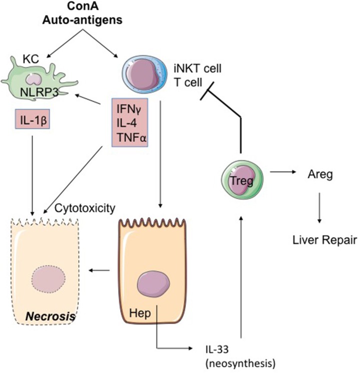
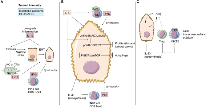
The pyrogenic property being the first activity described, members of the interleukin-1 superfamily (IL-1α, IL-1β, IL-18, and the newest members: IL-33, IL-36, IL-37, and IL-38) are now known to be involved in several inflammatory diseases such as obesity, atherosclerosis, cancer, viral and parasite infections, and auto-inflammatory syndromes as well as liver diseases. Inflammation processes are keystones of chronic liver diseases, of which the etiology may be viral or toxic, as in alcoholic or non-alcoholic liver diseases. Inflammation is also at stake in acute liver failure involving massive necrosis, and in ischemia-reperfusion injury in the setting of liver transplantation. The role of the IL-1 superfamily of cytokines and receptors in liver diseases can be either protective or pro-inflammatory, depending on timing and the environment. Our review provides an overview of current understanding of the IL-1 family members in liver inflammation, highlighting recent key investigations, and therapeutic perspectives. We have tried to apply the concept of trained immunity to liver diseases, based on the role of the members of the IL-1 superfamily, first of all IL-1β but also IL-18 and IL-33, in modulating innate lymphoid immunity carried by natural killer cells, innate lymphoid cells or innate T-αβ lymphocytes.

Keywords: inflammation; innate immunity; innate lymphoid cells; interleukin-1 family cytokines; invariant natural killer T-cells; liver diseases; natural killer cells; trained immunity.


Figures

Figure 1
Figure 1
IL-1 cytokine family intracellular and extracellular processing. (A) Intracellular processing. Caspase-1 cleavage from the NLRP3 inflammasome activates pro-IL-1β and pro-IL-18 and inactivates pro-IL-37, which is translocated in the nucleus. IL-1α and IL-33 are rapidly released by damaged necrotic cells in their functionally active full-length precursors and are inactivated by caspase-1, 3, and 7 cleavage. Intracellular processing of IL-36α, IL-36β, IL-36γ, and IL-38 are still unknown. (B) Extracellular processing. Epithelial cells, NK cells, macrophages, mast cells, and PMN secrete proteases to activate IL-1β, IL-18, IL-33, IL-36α, IL-36β, and IL-36γ. Unlike the intracellular processing, the cleavage of IL-33 and IL-37 results in a superactive form. NK, natural killer; PMN, polymorphonuclear neutrophils; PR3, proteinase 3.
Figure 2
Figure 2
IL-1α/β and IL-18 members are involved in ALF. (A) Acetaminophen-induced ALF. NAPQI (N-acetyl-p-benzoquinone-imine) is a toxic acetaminophen metabolite generated by hepatic cytochrome Cype2e1 and Cyp1a2. Under NAPQI action, injured and necrotic centrilobular hepatocytes secrete DAMP. Among them, ATP and TLR9 receptor ligand induce IL-1β and IL-18 production by Kupffer cells. Both cytokines drive NK and iNKT cell killing function through FasL and IFN-γ production. This inflammatory process can be blocked either by endogenous IL-18BP production by non-damaged hepatocytes, Kupffer and stellate cells, or by recombinant IL-18BP treatment. (B) Critical role of IL-18 in HAV fulminant hepatitis. HAV-infected hepatocytes produce IL-15 as well as PAMP and DAMP which, in turn, activate IL-18 production by Kupffer cells. IL-18 activates IFN-γ production by NK cells and CD8 T-cells with innate functions, hence promoting cytotoxicity toward infected hepatocytes. In healthy infected subjects (left), endogenous IL-18BP blocks the deleterious IL-18 action and modulates NK cell and probably CD8 T-cell activation. In IL-18BP deficient subjects (right), there is no inhibition of the deleterious effects of NK cell and CD8 T-cell activation. ALF, acute liver failure; KC, Kupffer cells; NK, natural killer; Hep, hepatocyte; bounded arrow, presumably yes.
Figure 3
Figure 3
Alcohol acts as an “exogenous signal” on Kupffer cells to activate the inflammasome NLRP3-caspase 1 and production of IL-1β in ALD. Alcohol decreases microRNA miR-148a, a negative regulator of TXNIP (thioredoxin-interacting protein), which is overexpressed during ALD. TXNIP activates NLRP3 inflammasome in hepatocytes and caspase 1-mediated pyroptosis, leading to IL-1β and IL-18 production. ATP and uric acid release by damaged hepatocytes after alcohol damage is a second “endogenous signal” leading directly and/or through Kupffer cell activation to inflammatory cytokine production. IL-1β and IL-18 recruit and activate iNKT cells that, in turn, induce PMN afflux and hepatic injury. IL-33 released by damaged and necrotic hepatocytes exacerbates iNKT cells and PMN recruitment and activation. ALD, alcoholic liver disease; KC, Kupffer cells; iNKT, invariant natural killer T-cells; PMN, polymorphonuclear neutrophils; Hep, hepatocyte.
Figure 4
Figure 4
IL-1β, together with TNFα, mediates low-grade inflammation during NAFLD. Free fatty acids, acting as DAMP, are capable together with TLR ligands of activating NLRP3 and NLRP6 inflammasomes, thereby inducing caspase-1 activation and release of IL-1α and IL-1β in hepatocytes and Kupffer cells. Adipocytes could be an important source of IL-1α and IL-β during NAFLD associated with obesity. Adipocyte-derived IL-1β has been shown to directly induce insulin resistance in hepatocyte development of insulin resistance in adipocytes enabling accumulation of lipids in the liver. IL-1β is also capable of reducing insulin receptor substrate-1 (IRS-1) expression dependently and independently of ERK activation and of inducing impairment in insulin signaling and action. IL-1β is released by hepatocytes through inflammasome activation and, in turn, amplifies inflammasome activation, TNFα, and IL-β release in Kupffer cells. Increased IL-1β and NLRP3 drive liver fibrosis in experimental models of NAFLD mice. IL-1β acts on LSEC to promote liver inflammation by upregulating ICAM-1 (intercellular adhesion molecule 1) expression, which stimulates neutrophil recruitment in the liver. IL-18 production negatively regulates NAFLD/NASH progression via modulation of the gut microbiota. NAFLD, Non-alcoholic fatty liver disease; KC, Kupffer cells; LSEC, liver sinusoidal endothelial cells.
Figure 5
Figure 5
The ConA model of hepatitis. The mitogen Concanavalin A (ConA) activates iNKT cells in a KC and iNKT cell-dependent mode. IL-4, IFNγ, and TNFα produced by hepatic activated iNKT cells activate the NLRP3 inflammasome leading to toxic IL-1β production by hepatocytes. iNKT cells could also induce IL-33 production by hepatocytes through TRAIL/DR5 interactions. Secreted IL-33 acts as a retro-negative regulator of iNKT cells by recruiting Treg. Indeed, Treg can restrain T-cell cytotoxicity and promote repair through signals like Areg. ConA, concanavalin A; Areg, Amphiregulin; KC, Kupffer cell; Hep, hepatocyte.
Figure 6
Figure 6
Role of IL-1 cytokine family members in the immunological history of the hepatocarcinoma. (A) IL-1β cytokine and trained immunity in initiation of hepatocarcinoma transformation. Trained immunity induced by metabolic syndrome or HFD/NAFLD leads to low-grade inflammation linked to IL-1β and other cytokine (as TNF-α) production. These cytokines induce hepatic fibrosis, exposing hepatocytes to moderate hypoxia. HIF-1α accumulation up-regulates IL-1β secretion in TAM/KC. Locally produced IL-1β induces or enhances epithelial-mesenchymal transition in cancer cells. At that time, iNKT cells and CD8 T-cells maintain the immunosurveillance of the HCC. (B) IL-37 driven-suppression of hepatocellular carcinoma. Hepatocytes and transformed hepatocytes produce IL-37, a cytokine with several cell targets. IL-37 suppresses hepatocellular carcinoma growth by converting pSmad3 signaling from JNK/pSmad3L/c-Myc oncogenic signaling into pSmad3C/P21 tumor-suppressive signaling. IL-37 inhibits the PI3K/AKT/mTOR pathway, inducing autophagy in hepatocellular carcinoma cells. IL-37 recruits cytotoxic anti-tumoral NK cells harboring the terminal differentiation marker CD57. At that time, IL-33 neosynthesis by hepatocarcinoma cells and hepatocytes could maintain the immunosurveillance by maintaining iNKT1 cell and T CD8 T-cell activation. (C) Immunosurveillance failure at the end-stage HCC. Sustained IL-33 production by tumoral cells can recruit Treg lymphocytes and promote development of iNKT2 CD4(+) cells, both leading to anti-tumoral immunosurveillance failure. Areg, Amphiregulin; EMT, epithelial-mesenchymal transition; HCC, hepatocellular carcinoma; KC, Kupffer cells; TAM, tumor-associated macrophages; bounded arrows, presumably yes.

References

    1. Menkin V. Studies on the isolation of the factor responsible for tissue injury in inflammation. Science. (1943) 97:165–7. 10.1126/science.97.2511.165 - DOI - PubMed
    1. Beeson PB. Temperature-elevating effect of a substance obtained from polymorphonuclear leucocytes. J Clin Invest. (1948) 27:524. - PubMed
    1. Atkins E, Wood WB, Jr. Studies on the pathogenesis of fever. II. Identification of an endogenous pyrogen in the blood stream following the injection of typhoid vaccine. J Exp Med. (1955) 102:499–516. 10.1084/jem.102.5.499 - DOI - PMC - PubMed
    1. Dayer JM. From supernatants to cytokines: a personal view on the early history of IL-1, IL-1Ra, TNF and its inhibitor in rheumatology. Arthritis Res Ther. (2018) 20:1–6. 10.1186/s13075-018-1607-y - DOI - PMC - PubMed
    1. Dinarello CA. Interleukin-1. Cytokine Growth Factor Rev. (1997) 8:253–65. 10.1016/S1359-6101(97)00023-3 - DOI - PubMed

Publication types

MeSH terms