. 2019 Aug 27:10:2020.
doi: 10.3389/fimmu.2019.02020. eCollection 2019.

MEK1/2 Inhibitors Unlock the Constrained Interferon Response in Macrophages Through IRF1 Signaling

Affiliations

MEK1/2 Inhibitors Unlock the Constrained Interferon Response in Macrophages Through IRF1 Signaling

Lei Yang et al. Front Immunol. .

Abstract

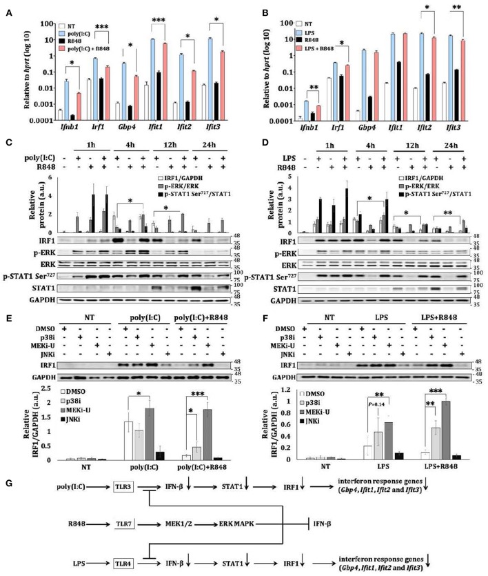
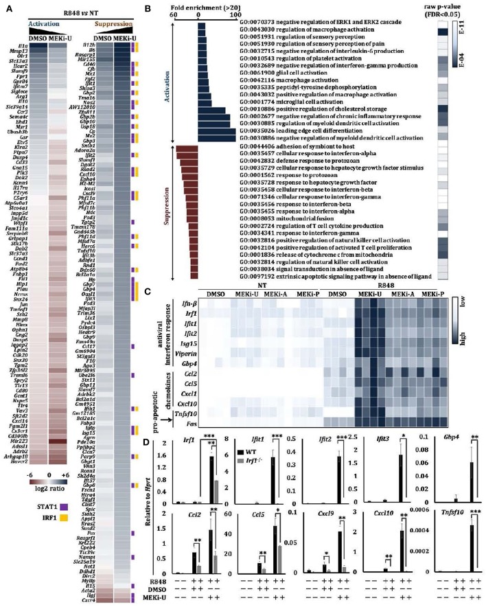
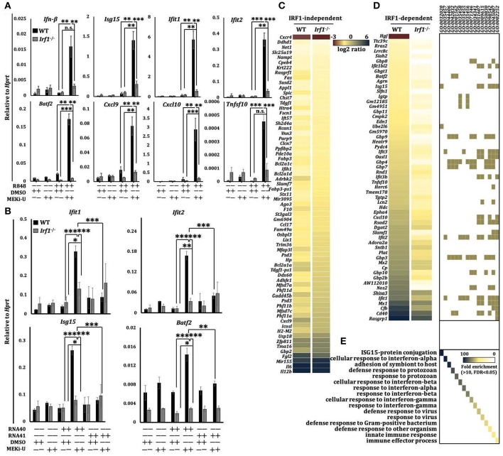
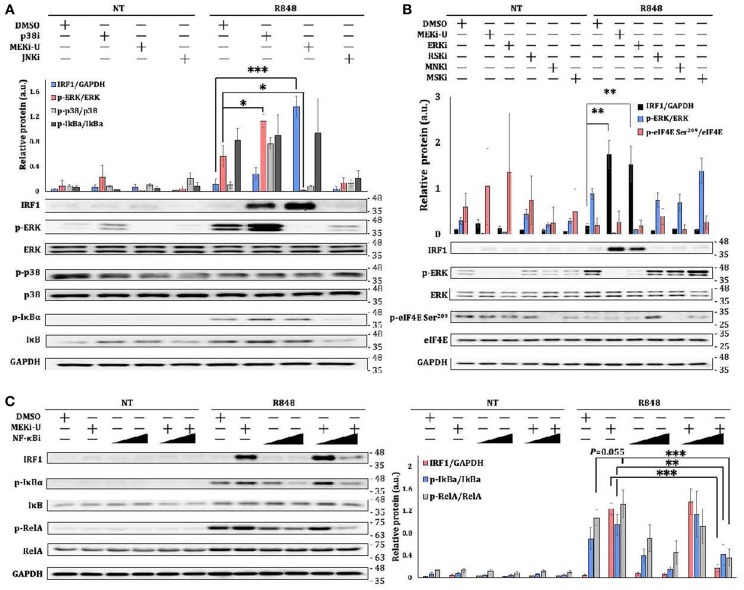
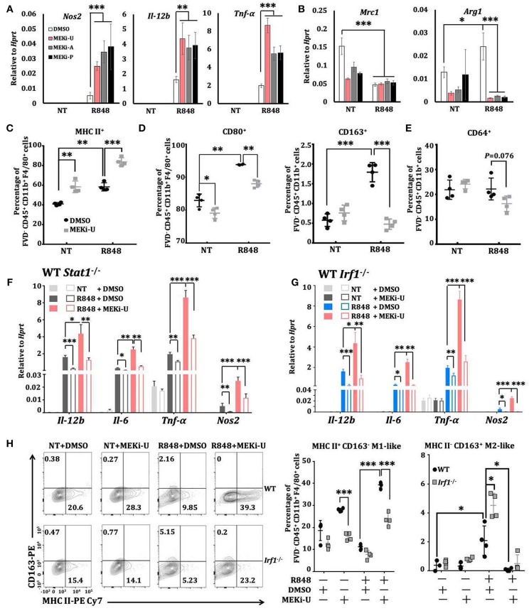
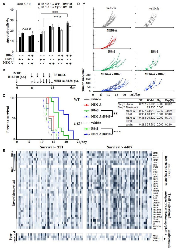
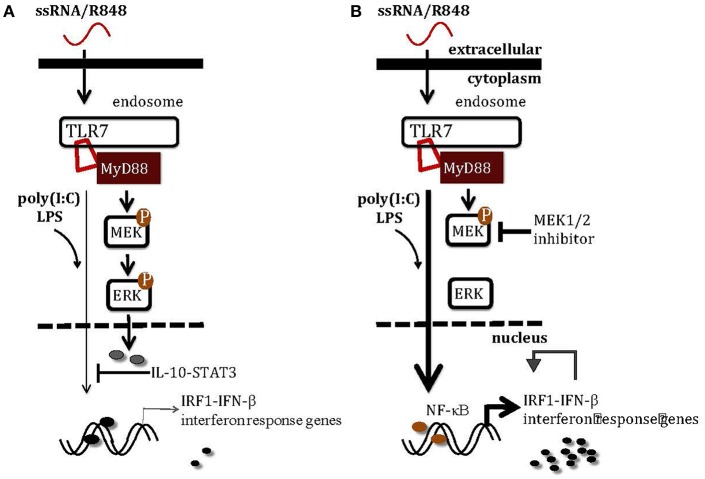
Macrophages are immune sentinels essential for pathogen recognition and immune defense. Nucleic acid-sensing toll-like receptors like TLR7 activate tailored proinflammatory and interferon responses in macrophages. Here we found that TLR7 activation constrained itself and other TLRs from inducing interferon response genes in macrophages through MAPK kinase 1/2 (MEK1/2)-dependent IRF1 inhibition. Downstream of the MEK1/2-ERK pathway, TLR7-activated macrophages induced interleukin-10 (IL-10), a signal transducer and activator of transcription 3 (STAT3) signaling axis, which constrained the expression of interferon response genes, immunomodulatory cytokines, and chemokines. Nevertheless, MEK1/2 inhibitors unlocked an IRF1-interferon signature response in an NF-κB-dependent manner. Deficiency in interferon regulatory factor 1 (Irf1) completely abrogated the interferon response and prevented the reprogramming of macrophages into an immunostimulatory phenotype. As a proof of concept, combination treatment with a TLR7 agonist and MEK1/2 inhibitor synergistically extended the survival of wild-type but not Irf1-deficient melanoma-bearing mice. In a retrospective study, higher expression of Irf1 and interferon response genes correlated with more favorable prognosis in patients with cutaneous melanoma. Our findings demonstrated how MEK1/2 inhibitor unlocks IRF1-mediated interferon signature response in macrophages, and the therapeutic potentials of combination therapy with MEK1/2 inhibitor and TLR7 agonist.

Keywords: IRF1; MAPK; MEK inhibitor; TLR7 agonist; interferon; macrophage; reprogramming.


Figures

Figure 1
Figure 1
TLR7 stimulation constrains expression of interferon response genes during TLR crosstalk in macrophages. (A,B) qRT-PCR analysis of Ifn-β, Irf1, Gbp4, Ifit1, Ifit2, and Ifit3 mRNA expression in BMDM stimulated as indicated for 12 h. Data are means ± SD from 4 experiments. (C,D) Immunoblot analysis and quantitative densitometry of IRF1, total and p-ERK, total and p-STAT1 in BMDM stimulated for indicated time intervals. Blots are representative of 3 or 4 experiments. Quantified data are means ± SD from all experiments. (E,F) Immunoblot analysis and quantitative densitometry of IRF1 in stimulated BMDMs stimulated with TLR3 agonist poly(I:C) and TLR7 agonist R848 (E), or with TLR4 agonist LPS and R848 (F) for 12 h in the presence or absence of indicated MAPK inhibitors. Blots are representative of 4 or 5 experiments. Molecular weight (kDa) markers are indicated on the right side of the blots. Quantified data are means ± SD from all experiments. (G) Schematic illustration of TLR7-specific suppression on TLR3- and TLR4- induced interferon response and induction of intetferon response genes like Irf1. TLR7 also activates the MEK1/2-ERK MAPK pathway to limit itself from inducing interferon response. *P < 0.05, **P < 0.01, and ***P < 0.001 by unpaired Welch's t-test.
Figure 2
Figure 2
MEK1/2 inhibitor synergizes with TLR7 agonist to unlock an interferon signature response. (A,B) Microarray analysis of BMDMs treated with TLR7 agonist R848 in the presence or absence of MEKi-U for 6 h (see also Figure S2A). Heat map (A) and Gene set enrichment analysis (B) of differentially expressed genes are from 2 independent experiments. Differentially expressed genes interacting with STAT1 and IRF1 were labeled with purple and orange squares based on STRING PPI test. (C) Heat map showing qRT-PCR analysis of the mRNA expression of selected interferon response genes, chemokines, and pro-apoptotic genes. Data are from 4 independent experiments. (D) qRT-PCR analysis of interferon response genes, chemokines, and pro-apoptotic genes in WT and Stat1−/− BMDMs treated with TLR7 agonist R848 in the presence or absence of MEKi-U for 6 h. Data are means ± SD from 4 independent experiments. *P < 0.05, **P < 0.01, and ***P < 0.001 by unpaired Welch's t-test.
Figure 3
Figure 3
Unlocked interferon signature response is IRF1-dependent. (A) qRT-PCR analysis of mRNA expression of the indicated interferon response genes in WT and Ifit1−/− BMDM stimulated with TLR7 agonist R848 for 6 h in the presence or absence of MEK1/2 inhibitor (MEKi-U). Data are means ± SD from 3 to 5 independent experiments. (B) qRT-PCR analysis of Ifil1, Ifit2, Isgl5, and Batj2 mRNA expression in BMDM stimulated with RNA40 or RNA41 for 6 h in the presence or absence of MEKi-U. Data are means ± SD from 4 independent experiments. (C–E) Microarray analysis of mRNA expression in WT and Ifit1−/− BMDM stimulated with TLR7 agonist R848 for 6 h in the presence or absence of MEKi-U. Heat map of IRF1-independent (C) or dependent (D) genes clustered using 1.5-fold difference cutoff. Gene set enrichment analysis of differentially expressed genes (E) are from all replicates. Quantified data are means ± SD from all experiments. *P < 0.05, **P < 0.01, and ***P < 0.001 by unpaired Welch's t-test. n.s., not significant.
Figure 4
Figure 4
IRF1 is unlocked through the activation of NF-κB pathway. (A–C) Immunoblot analysis and quantitative densitometry of the indicated proteins in lysates from BMDM activated with R848 in the presence or absence of MEKi-U, p38 MAPK inhibitor p38i, or JNK MAPK inhibitor JNKi (A), or MEKi-U, ERK inhibitor (ERKi), RSK inhibitor (RSKi), MNK inhibitor (MNKi), or MSK inhibitor (MSKi) (B), or MEKi-U, and NF-κB inhibitor NF-κBi (C) for 12 h (A,C) or 8 h (B). Blots are representative of 4 independent experiments. Molecular weight (kDa) markers are indicated on the right side of the blots. Quantified data are means ± SD from all experiments. *P < 0.05, **P < 0.01, and ***P < 0.001 by unpaired Welch's t-test. n.s., not significant.
Figure 5
Figure 5
MEK1/2 pathway constrains IRF1-mediated interferon signature response through the IL-10 signaling. (A) ELISA analysis of IL-10 secretion from BMDM stimulated as indicated. Data are means ± SD pooled from 3 experiments. (B) qRT-PCR analysis of Il-10 mRNA expression in BMDM stimulated with R848 for 6 h in the presence or absence of three MEK1/2 inhibitors. Data are means ± SD pooled from 4 experiments. (C) Immunoblot analysis and quantitative densitometry of IRF1, total, and p-STAT3 abundance in lysates of BMDM treated with R848 for 8 h in the presence or absence of MEKi-U, anti-IL-10R antibody, and IgG isotype control. Blots are representative of 4 independent experiments. Molecular weight (kDa) markers are indicated on the right side of the blots. Quantified data are means ± SD from all experiments. (D) qRT-PCR analysis of mRNA expression of the indicated genes in BMDM cells stimulated with R848 and recombinant IL-10 (20 ng/ml) for 6 h in the presence or absence of MEKi-U. Data are means ± SD pooled from 4 experiments. *P < 0.05, **P < 0.01, and ***P < 0.001 by one-way ANOVA (B) or unpaired Welch's t-test (C,D).
Figure 6
Figure 6
Macrophages are reprogrammed toward a predominantly M1-like phenotype through IRF1. (A,B) qRT-PCR analysis of Nos2, Il-12b, and Tnfα (A) or Mrc1 and Arg1 (B) mRNA expression in BMDMs activated with TLR7 agonist R848 for 6 h in the presence or absence of different MEK1/2 inhibitors. Data are means ± SD pooled from 4 experiments. (C–E) Flow cytometry analysis of MHC II (C), CD80 and CD163 (D), and CD64 (E) surface expression on BMDMs stimulated with R848 for 24 h in the presence or absence of MEKi-U. Data with means ± SD are from 4 independent experiments. (F,G) qRT-PCR analysis of the indicated mRNAs expressed in WT and Stat1−/− (F) or Irf1−/− (G) BMDMs activated with TLR7 agonist R848 for 6 h in the presence or absence of different MEK1/2 inhibitors. Data are means± SD pooled from 4 experiments. (H) Flow cytometry analysis of MHC II+ CD163 M1 and MHC II CD163+ M2 phenotypes in WT and Irf1−/−mice BMDM stimulated with R848 for 24 h in the presence or absence of MEKi-U. Contour plots are representative of 4 independent experiments. Quantified% positive data with means ± SD are from all experiments. *P < 0.05, **P < 0.01, and ***P < 0.001 by one-way ANOVA (A,B) with Bonferroni's correction (F,G) or unpaired Welch's t-test (C–H).
Figure 7
Figure 7
Combination therapy with MEK1/2 inhibitor and TLR7 agonist reduces melanoma progression in vivo. (A) Flow cytometry analysis of B16F10 apoptosis cultured alone or co-cultured with WT or Irf1−/− BMDM. Cells were stimulated with R848 in the presence or absence of MEKi-U for 48 h. Data are means ± SD from 4 independent experiments. (B–D) WT or Irf1−/− mice bearing subcutaneous (s.c.) B16F10 melanoma tumors were treated with MEKi-A or intratumoral (i.t.) R848 or both as indicated in (B). (C,D) Survival analysis (C) and tumor growth (D) of WT or Irf1−/− melanoma-bearing mice treated with MEKi-A and/or R848. Data from at least 9 mice/group are pooled from 2 to 3 independent experiments. (E) Heat map showing mRNA expression of 36 genes identified with significant associations between their expression levels and patient survival. Data presented are from individuals with overall survival shorter than 321 days (n = 50) or longer than 4,407 days (n = 50). *P < 0.05, **P < 0.01, ***P < 0.001 by unpaired Welch's t-test (A), Cox regression analysis (C), or log-rank test (C,E).
Figure 8
Figure 8
A schematic illustration of how MEK1/2 pathway modulates the interferon responses in macrophages. (A) TLR3 activation by poly(I:C) and TLR4 activation by LPS induce scarce levels of Irf1 and interferon response genes, at least in part due to the inhibitory effects of MEK1/2-ERK-IL-10-STAT3 signaling. (B) Inhibition of MEK1/2 pathway using MEK1/2 inhibitor blocks activation of ERK-mediated inhibitory signaling, unlocking the IRF1-IFN-β-interferon signature response through the NF-κB pathway. The combination therapy with TLR7 agonist and MEK1/2 inhibitor may greatly enhance the anti-tumor monotherapies with either TLR7 agonist or MEK1/2 inhibitor by boosting the interferon signature response.

Similar articles

Cited by

References

    1. Takeuchi O, Akira S. Pattern recognition receptors and inflammation. Cell. (2010) 140:805–20. 10.1016/j.cell.2010.01.022 - DOI - PubMed
    1. Brubaker SW, Bonham KS, Zanoni I, Kagan JC. Innate immune pattern recognition: a cell biological perspective. Annu Rev Immunol. (2015) 33:257–90. 10.1146/annurev-immunol-032414-112240 - DOI - PMC - PubMed
    1. Schlee M, Hartmann G. Discriminating self from non-self in nucleic acid sensing. Nat Rev Immunol. (2016) 16:566–80. 10.1038/nri.2016.78 - DOI - PMC - PubMed
    1. Heil F, Hemmi H, Hochrein H, Ampenberger F, Kirschning C, Akira S, et al. . Species-specific recognition of single-stranded RNA via toll-like receptor 7 and 8. Science. (2004) 303:1526–9. 10.1126/science.1093620 - DOI - PubMed
    1. Schneider WM, Chevillotte MD, Rice CM. Interferon-stimulated genes: a complex web of host defenses. Annu Rev Immunol. (2014) 32:513–45. 10.1146/annurev-immunol-032713-120231 - DOI - PMC - PubMed

Publication types

MeSH terms