. 2019 Aug 22:10:2021.
doi: 10.3389/fimmu.2019.02021. eCollection 2019.

Interleukin-1/-33 Signaling Pathways as Therapeutic Targets for Endometriosis

Affiliations

Interleukin-1/-33 Signaling Pathways as Therapeutic Targets for Endometriosis

Toru Kato et al. Front Immunol. .

Abstract

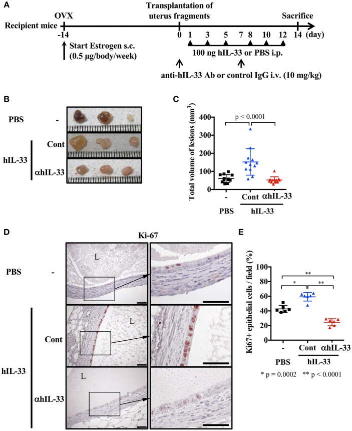
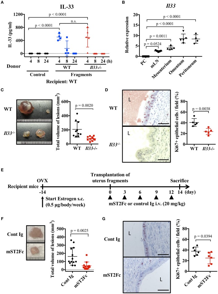
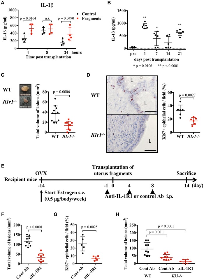
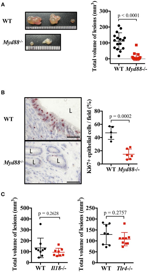
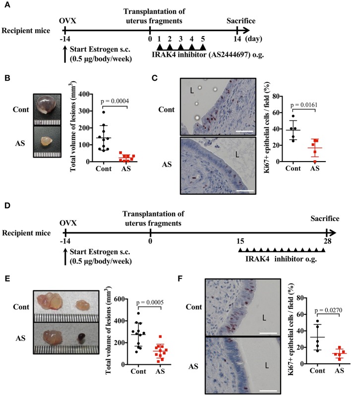
Endometriosis is an estrogen-dependent disease with symptoms of dysmenorrhea, chronic pain, and infertility that affects 6-10% of women of reproductive age. Medical or surgical therapy, such as administration of an anti-gonadotropin or ovarian cystectomy, provide effective pain relief. However, neither therapy can be used for patients wishing to become pregnant. Despite the high morbidity, the pathogenesis of endometriosis has not been well-elucidated. Several inflammatory cytokines are reported to participate in the onset of endometriosis. Here, we examined the role of interleukin (IL)-1/IL-33 signaling in the development of endometriosis using a mouse model of endometriosis. Endometriotic lesion volume was significantly reduced in Il33-/- and Il1r1-/- mice, and almost completely suppressed in Myd88-/- mice. Mice intraperitoneally administered with an antibody against IL-1 receptor 1 (IL-1R1) or IL-33 developed limited endometriotic lesions. Oral administration of an inhibitor against IL-1R-associated kinase 4 (IRAK4), a downstream signal molecule of MyD88, also suppressed lesion formation. Furthermore, even after the development of cystic lesions the IRAK4 inhibitor prevented the enlargement of lesions. These treatments all significantly reduced cellular proliferation, shown by decreased Ki-67 expression. These results reveal that IL-1/IL-1R1, IL-33/IL-33R and associated downstream signaling molecules are involved in the pathogenesis of endometriosis, and may provide novel therapeutic targets for endometriosis.

Keywords: IL-1R1; IRAK4; MyD88; ST2; estrogen.


Figures

Figure 1
Figure 1
Exogenous IL-33 exacerbates endometriosis. (A) Experimental workflow. Wild type BALB/c mice were ovariectomized (OVX) and administered estrogen subcutaneously (s.c.) for 2 weeks before transplantation of uterine fragments. After transplantation, mice received PBS or recombinant human IL-33 intraperitoneally (i.p.) at day 1, 3, 5, 8, 10, and 12. For the neutralization experiment, IL-33-treated mice were intravenously (i.v.) injected with control IgG (Cont) or anti-hIL-33 Ab (αhIL-33) at day 0 and 7. (B) Representative endometriosis lesions from each mouse. (C) Total volume of the lesions (n = 12 in each group). Pooled data from two independent experiments are shown (mean ± SD). (D) Immunohistological staining of Ki-67; Brown. L, lumen, Scale bar: 50 μm. (E) The proportion of Ki-67 positive epithelial cells lining the lumen of the cyst wall (n = 6, mean ± SD). Statistical analyses were performed using a one-way ANOVA with Tukey's post-hoc tests (C,E).
Figure 2
Figure 2
Endogenous IL-33 contributes to lesion formation in endometriosis. (A) Concentration of IL-33 in peritoneal lavage fluid. Wild type (WT) BALB/c mice were injected with PBS or uterine fragments transplanted from WT or Il33−/− mice. Peritoneal lavage fluids were collected at indicated time points. (B) Il33 mRNA expression in intraperitoneal tissues was measured by real-time RT-PCR. PC: peritoneal cells in peritoneal lavage fluid, mLN: mesenteric lymph node. (C) Mouse endometriosis model using WT mice or Il33−/− mice. (Left) Representative endometriosis lesions from each mouse. (Right) Total volume of the lesions (n = 10 in each group). Pooled data from two independent experiments are shown (mean ± SD). (D) (Left) Immunohistological staining of Ki-67; Brown. L: lumen, Scale bar: 50 μm. Low-power image was shown in Supplementary Figure 2A. (Right) The proportion of Ki-67 positive cells in epithelium (n = 5, mean ± SD). (E) Mouse endometriosis model performed with WT BALB/c mice. After uterine fragment transplantation, mice were treated with mST2Fc or control (Cont) Ig at the indicated days. (F) (Left) Representative endometriosis lesion. (Right) Total volume of the lesions (cont; n = 13, mST2Fc; n = 12). Pooled data from two independent experiments are shown (mean ± SD). (G) (Left) Immunohistological staining of Ki-67. Scale bar: 50 μm. Low-power image was shown in Supplementary Figure 2B. (Right) The proportion of Ki-67 positive cells in epithelium (n = 6, mean ± SD). Statistical analyses were performed using a one-way ANOVA with Tukey's post-hoc tests (A,B) or a Student's t-tests (C,D,F,G).
Figure 3
Figure 3
IL-1 exacerbates endometriosis lesions. (A,B) Concentration of IL-1β in peritoneal lavage fluids (A, n = 4), or serum (B, n = 6). Wild type (WT) BALB/c mice were injected with PBS or transplanted with uterine fragments from WT mice. Peritoneal lavage fluids or serum were collected at indicated time points. Statistical analyses were performed using a two-way ANOVA with Holm-Sidak post-hoc tests (A) or a one-way ANOVA with Tukey's post-hoc tests. (C) Mouse endometriosis model using WT C57BL/6 mice or Il1r1−/− mice. (Left) Representative endometriosis lesion. (Right) Total volume of the lesions (WT; n = 11, Il1r1−/−; n = 9). Pooled data from two independent experiments are shown (mean ± SD). (D) (Left) Immunohistological staining of Ki-67. Scale bar: 50 μm. Low-power image was shown in Supplementary Figure 2C. (Right) Proportion of Ki-67 positive cells in epithelium (n = 6, mean ± SD). (E) Mouse endometriosis model performed with WT BALB/c mice. One day before and 4 and 8 days after uterine fragment transplantation, mice were treated with anti-IL-1R1 Ab (αIL-1R1) or control (Cont) Ab. (F) Total volume of the lesions (cont; n = 9, anti-IL-1R1; n = 8). Pooled data from two independent experiments are shown (mean ± SD). (G) Immunohistological staining of Ki-67 was performed. Proportion of Ki-67 positive cells in epithelium are shown (n = 5, mean ± SD). Statistical analyses were performed using a Student's t-tests (C,D,F,G). (H) Mouse endometriosis model performed as in (E) with WT and Il33−/− mice. Total volume of the lesions (WT: cont; n = 9, Il33−/−: cont; n = 9, anti-IL-1R1; n = 8). Pooled data from two independent experiments are shown (mean ± SD). Statistical analyses were performed using a one-way ANOVA with Tukey's post-hoc tests.
Figure 4
Figure 4
MyD88 signaling is essential for endometriotic lesion formation. (A) Mouse endometriosis model performed with wild type (WT) BALB/c mice or Myd88−/− mice. (Left) Representative endometriosis lesions. (Right) Total volume of the lesions (n = 20). Pooled data from three independent experiments are shown (mean ± SD). (B) (Left) Immunohistological staining of Ki-67. Scale bar: 50 μm. Low-power image was shown in Supplementary Figure 2D. (Right) Proportion of Ki-67 positive cells in epithelium (WT; n = 5, Myd88−/−; n = 6, mean ± SD). (C) Mouse endometriosis model performed with WT, Il18−/−, and Tlr4−/− BALB/c mice. Total volume of the lesions (WT vs. Il18−/−, WT: n = 9, Il18−/−: n = 8; WT vs. Tlr4−/−, WT: n = 8, Tlr4−/−: n = 9). Pooled data from two independent experiments are shown (mean ± SD). Statistical analyses were performed using a Student's t-tests.
Figure 5
Figure 5
Blockade of MyD88 signaling suppresses endometriosis. (A) Mouse endometriosis model performed with WT BALB/c mice. After uterine fragment transplantation, mice were treated with AS2444697 (AS) or sorbent (cont) at indicated time points. o.g., oral gavage. (B) Total volume of the lesions (cont; n = 10, AS; n = 8). Pooled data from two independent experiments are shown (mean ± SD). (C) (Left) Immunohistological staining of Ki-67. Scale bar: 50 μm. Low-power image was shown in Supplementary Figure 2E. (Right) Proportion of Ki-67 positive cells in epithelium (n = 5, mean ± SD). (D) Mouse endometriosis model performed with WT BALB/c mice as in A. Administration of inhibitor was started at 2 weeks after transplantation. (E) Total volume of the lesions (cont; n = 12, AS; n = 11). Pooled data from two independent experiments are shown (mean ± SD). (F) (Left) Immunohistological staining of Ki-67. Scale bar: 50 μm. Low-power image was shown in Supplementary Figure 2F. (Right) Proportion of Ki-67 positive cells in epithelium (n = 5, mean ± SD). Statistical analyses were performed using a Student's t-tests.

Similar articles

Cited by

References

    1. Giudice LC. Clinical practice. Endometriosis. N Engl J Med. (2010) 362:2389–98. 10.1056/NEJMcp1000274 - DOI - PMC - PubMed
    1. Eskenazi B, Warner ML. Epidemiology of endometriosis. Obstet Gynecol Clin North Am. (1997) 24:235–58. 10.1016/S0889-8545(05)70302-8 - DOI - PubMed
    1. Kvaskoff M, Horne AW, Missmer SA. Informing women with endometriosis about ovarian cancer risk. Lancet. (2017) 390:2433–4. 10.1016/S0140-6736(17)33049-0 - DOI - PubMed
    1. Sampson JA. Metastatic or embolic endometriosis, due to the menstrual dissemination of endometrial tissue into the venous circulation. Am J Pathol. (1927) 3:93–110.43. - PMC - PubMed
    1. Iwabe T, Harada T, Tsudo T, Nagano Y, Yoshida S, Tanikawa M, et al. . Tumor necrosis factor-alpha promotes proliferation of endometriotic stromal cells by inducing interleukin-8 gene and protein expression. J Clin Endocrinol Metab. (2000) 85:824–9. 10.1210/jcem.85.2.6335 - DOI - PubMed

Publication types