

Bta-miR-10b Secreted by Bovine Embryos Negatively Impacts Preimplantation Embryo Quality
- PMID: 31507632
- PMCID: PMC6713719
- DOI: 10.3389/fgene.2019.00757
Bta-miR-10b Secreted by Bovine Embryos Negatively Impacts Preimplantation Embryo Quality
Abstract
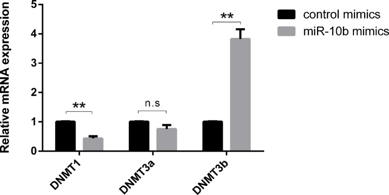
In a previous study, we found miR-10b to be more abundant in a conditioned culture medium of degenerate embryos compared to that of blastocysts. Here, we show that miR-10b mimics added to the culture medium can be taken up by embryos. This uptake results in an increase in embryonic cell apoptosis and aberrant expression of DNA methyltransferases (DNMTs). Using several algorithms, Homeobox A1 (HOXA1) was identified as one of the potential miR-10b target genes and dual-luciferase assay confirmed HOXA1 as a direct target of miR-10b. Microinjection of si-HOXA1 into embryos also resulted in an increase in embryonic cell apoptosis and downregulation of DNMTs. Cell progression analysis using Madin-Darby bovine kidney cells (MDBKs) showed that miR-10b overexpression and HOXA1 knockdown results in suppressed cell cycle progression and decreased cell viability. Overall, this work demonstrates that miR-10b negatively influences embryo quality and might do this through targeting HOXA1 and/or influencing DNA methylation.
Keywords: DNA methylation; HOXA1; apoptosis; bovine embryos; miR-10b; secreted miRNAs.
Figures


Similar articles
-
Bovine Embryo-Secreted microRNA-30c Is a Potential Non-invasive Biomarker for Hampered Preimplantation Developmental Competence.Front Genet. 2019 Apr 5;10:315. doi: 10.3389/fgene.2019.00315. eCollection 2019. Front Genet. 2019. PMID: 31024625 Free PMC article.
-
ERRATUM.Oncol Res. 2020 Sep 1;28(4):447-449. doi: 10.3727/096504020X15970683241756. Oncol Res. 2020. PMID: 32859282 Free PMC article.
-
MicroRNA-146b negatively affects bovine embryo development and quality.Reproduction. 2023 Dec 1:REP-23-0155. doi: 10.1530/REP-23-0155. Online ahead of print. Reproduction. 2023. PMID: 38063339
-
MiR-10b regulates the proliferation and apoptosis of pediatric acute myeloid leukemia through targeting HOXD10.Eur Rev Med Pharmacol Sci. 2018 Nov;22(21):7371-7378. doi: 10.26355/eurrev_201811_16275. Eur Rev Med Pharmacol Sci. 2018. PMID: 30468483
-
MicroRNA-99a-5p alleviates atherosclerosis via regulating Homeobox A1.Life Sci. 2019 Sep 1;232:116664. doi: 10.1016/j.lfs.2019.116664. Epub 2019 Jul 17. Life Sci. 2019. PMID: 31325426
Cited by
-
Hatching is modulated by microRNA-378a-3p derived from extracellular vesicles secreted by blastocysts.Proc Natl Acad Sci U S A. 2022 Mar 22;119(12):e2122708119. doi: 10.1073/pnas.2122708119. Epub 2022 Mar 17. Proc Natl Acad Sci U S A. 2022. PMID: 35298333 Free PMC article.
-
Bta-miR-665 improves bovine blastocyst development through its influence on microtubule dynamics and apoptosis.Front Genet. 2024 Oct 16;15:1437695. doi: 10.3389/fgene.2024.1437695. eCollection 2024. Front Genet. 2024. PMID: 39479397 Free PMC article.
-
Murine Blastocysts Release Mature MicroRNAs Into Culture Media That Reflect Developmental Status.Front Genet. 2021 May 28;12:655882. doi: 10.3389/fgene.2021.655882. eCollection 2021. Front Genet. 2021. PMID: 34122510 Free PMC article.
-
tRNAGlu-derived fragments from embryonic extracellular vesicles modulate bovine embryo hatching.J Anim Sci Biotechnol. 2024 Mar 1;15(1):23. doi: 10.1186/s40104-024-00997-7. J Anim Sci Biotechnol. 2024. PMID: 38424649 Free PMC article.
-
MicroRNA-148b secreted by bovine oviductal extracellular vesicles enhance embryo quality through BPM/TGF-beta pathway.Biol Res. 2024 Mar 23;57(1):11. doi: 10.1186/s40659-024-00488-z. Biol Res. 2024. PMID: 38520036 Free PMC article.
References
LinkOut - more resources
Full Text Sources
Research Materials