. 2019 Sep 5:8:29.
doi: 10.1186/s40035-019-0166-8. eCollection 2019.

Xenografting of human umbilical mesenchymal stem cells from Wharton's jelly ameliorates mouse spinocerebellar ataxia type 1

Affiliations

Xenografting of human umbilical mesenchymal stem cells from Wharton's jelly ameliorates mouse spinocerebellar ataxia type 1

Pei-Jiun Tsai et al. Transl Neurodegener. .

Abstract

Background: Spinocerebellar ataxia type 1 (SCA1) is an autosomal dominant neurodegenerative disorder caused by the expansion of CAG repeats in ATXN1 gene resulting in an expansion of polyglutamine repeats in the ATXN1 protein. Unfortunately, there has yet been any effective treatment so far for SCA1. This study investigated the feasibility of transplanting human umbilical mesenchymal stem cells (HUMSCs) into transgenic SCA1 mice containing an expanded uninterrupted allele with 82 repeats in the ATXN1-coding region.

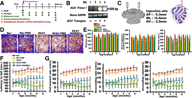
Methods: 106 human umbilical mesenchymal stem cells were transplanted into the cerebella at 1 month of age.

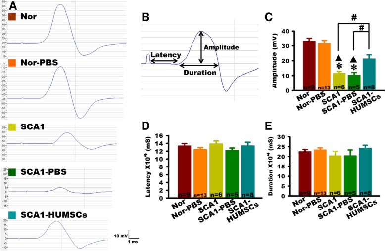
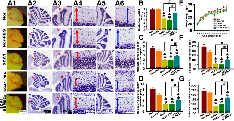
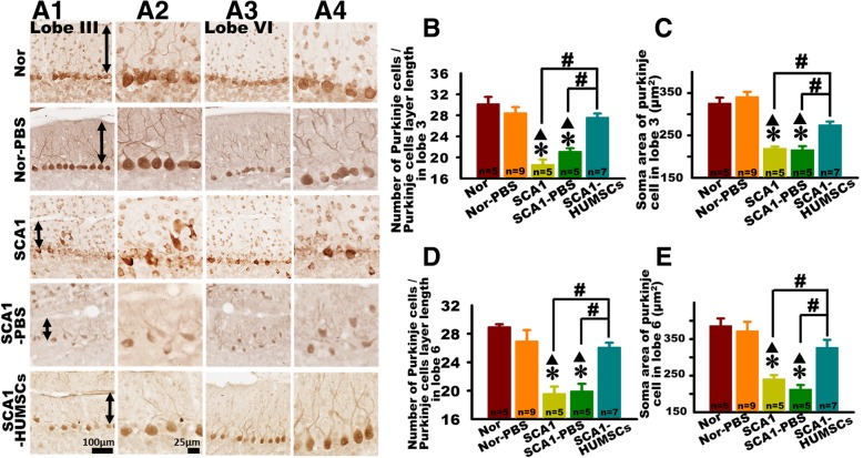
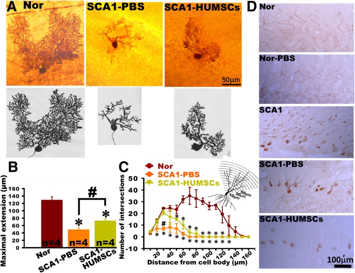
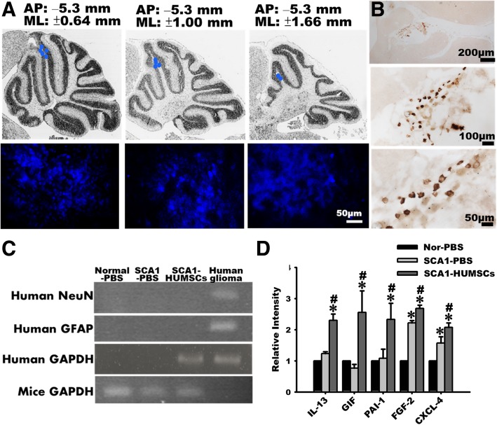
Results: HUMSCs displayed significant ameliorating effects in SCA1 mice in terms of motor behaviors in balance beam test and open field test as compared with the untransplanted SCA1 mice. HUMSCs transplantation effectively reduced the cerebellar atrophy, salvaged Purkinje cell death, and alleviated molecular layer shrinkage. Electrophysiological studies showed higher amplitudes of compound motor action potentials as indicated by increasing neuronal-muscular response strength to stimuli after stem cell transplantation. At 5 months after transplantation, HUMSCs scattering in the mice cerebella remained viable and secreted cytokines without differentiating into neuronal or glia cells.

Conclusions: Our findings provide hope for a new therapeutic direction for the treatment of SCA1.

Keywords: Cell transplantation; SCA1; Umbilical mesenchymal stem cells.


Conflict of interest statement

Competing interestsThe authors declare that they have no competing interests.

Figures

Fig. 1
Fig. 1
HUMSCs transplantation ameliorated motor behavior deterioration in SCA1 mice. a Scheme of experimental procedures. b Genotyping of SCA1 transgene. SCA1 Primer 1 set amplified a 500-bp amplicon. Lanes 3 and 4 show the presence of the SCA1 transgene. c The sketch and HE-stained picture show the injection site of HUMSCs. Open field tests revealed that HUMSCs transplants improved total distance traveled, rearing frequency, and rearing time in the SCA1-HUMSCs group mice (d and e). The group of SCA1-HUMSCs received, at 1 month of age, the HUMSCs transplantation which ameliorated the significant shortening of the latency to fall in the rotarod test (f), flat-forward, descending, and ascending speeds in the balance beam test (g) suffered by the SCA1 mice. * p < 0.05 compared with the Normal group of the same age. ▲ p < 0.05 compared with the Normal-PBS group of the same age. # p < 0.05 compared with the SCA1-HUMSCs group of the same age
Fig. 2
Fig. 2
HUMSCs transplantation improved the amplitude of the compound motor action potentials in SCA1 mice. a Representative recordings of compound motor action potentials generated by the gastrocnemius muscles at 6 months of age for Normal, Normal-PBS, SCA1, SCA1-PBS, and SCA1-HUMSCs mice, showing markedly depressed action potentials in the SCA1 and SCA1-PBS groups and partial recovery by HUMSC transplantation. b Parameters of a schematic compound motor action potential assessed in the study. Quantifications of the mean amplitude, latency, and duration of the compound motor action potentials indicated, in the 6 months old SCA1 and SCA1-PBS mice, significant amplitude depressions which were partially recovered in the SCA1-HUMSCs group (c). However, no statistical differences were found among all the groups with respect to latency (d) and duration (e) recorded. * p < 0.05 compared with the Normal group. ▲ p < 0.05 compared with the Normal-PBS group. # p < 0.05 compared with the SCA1-HUMSCs group
Fig. 3
Fig. 3
HUMSCs transplantation alleviated molecular layer atrophy in six-month-old SCA1 mice. Six-month-old mice were sacrificed to examine alterations within cerebellar sagittal sections (a1). The areas of cerebellar sagittal section were quantified. HUMSCs transplantation reduced the shrinkage of cerebellum in SCA1 mice (b). All groups were examined, photomicrographs of entire sagittal sections were stained with Cresyl violet (a2-a6), lobule III at low (red arrow, a2 and a3) and high (a4) magnification, and lobule VI at low (blue arrow, a2 and a5) and high (a6) magnification were shown. In lobules III and VI, significant reductions were found in the molecular layer areas (c and d) and thickness (f and g) in the six-month-old SCA1 and SCA1-PBS groups. HUMSCs transplants could partially mitigate these reductions in SCA1-HUMSCs mice. There were no significant differences in the body weight among mice in all groups at the same ages (e). * p < 0.05 compared with the Normal group. ▲p < 0.05 compared with the Normal-PBS group. # p < 0.05 compared with the SCA1-HUMSCs group
Fig. 4
Fig. 4
HUMSCs transplantation alleviated Purkinje cell damage SCA1 mice. Damages to the Purkinje cells in the SCA1 mice were assessed by sacrificing six-month-old SCA1 mice and observing, under optical microscope, Purkinje cells in lobules III and VI in tissue slices following immunohistochemical staining with anti-calbindin antibodies. Presented are representative photomicrographs of tissue slices of lobules III (a1 and a2) and VI (a3 and a4) in mice at 6 months of age in all groups. Purkinje cell number per unit length (b and d) and cell body area (c and e) in lobules III and VI were quantified. At 6 months of age, significant decreases were observed in both cell number and cell body area in the groups of SCA1 and SCA1-PBS. Transplantation of HUMSCs salvaged such losses (b-e). * p < 0.05 compared with the Normal group. ▲ p < 0.05 compared with the Normal-PBS group. # p < 0.05 compared with the SCA1-HUMSCs group
Fig. 5
Fig. 5
HUMSCs transplantation alleviated decline in dendritic branching in Purkinje cells. Cerebellar atrophy, as assessed by decline in dendritic branching in the Purkinje cells, was followed by morphological observations in cerebellar slices (300 μm thick) from six-month-old mice following intracellular biotin injection and immunostaining with anti-biocytin antibodies. Presented are representative photomicrographs and corresponding schematic drawings of cerebellar slices from mice in the groups of Normal, SCA1-PBS and SCA1-HUMSCs (a). Centered at the soma body, concentric rings with a radial distance of 10 μm between circles were used for quantification of the number of intersections. Quantifications using Sholl’s analysis indicated that HUMSCs transplantation partially preserved Purkinje cell extensions (b) and ameliorated shrinkage of intersections in the proximal regions (c) in SCA1 mice. Cerebellar sections were stained with an ATXN1 antibody. Presence of robust staining of ATXN1 protein in nuclei were found in the groups SCA1 and SCA1-PBS mice but not in the Normal, Normal-PBS, and SCA1-HUMSCs mice (d). * p < 0.05 compared with the Normal group. # p < 0.05 compared with the SCA1-HUMSCs group
Fig. 6
Fig. 6
Transplanted HUMSCs survived in the grafted-cerebellum and did not differentiate into neuronal cells. HUMSCs were labeled with the blue fluorescent nuclear staining bisbenzamide 48 h prior to transplantation and their viability and distribution were followed when the mice were sacrificed at 6 months of age. Blue fluorescent cell clusters were identified at lateral levels of bregma 1.2 mm, 2.1 mm, and 3.0 mm, with upper panel displaying the relative sites, lower panel showing the fluorescence photographs (a). Samples from six-month-old SCA1-HUMSCs mice were immunostained with antibody against human specific nuclei antigen (b). c RT-PCR was further applied to detect human NeuN and GFAP mRNA expressions in SCA1 mice. Human NeuN and GFAP mRNAs were not detectable in the SCA1-HUMSCs group. Human glioma cells were used as a positive control of human NeuN and GFAP mRNAs assay. d The expression of 174 human cytokines in mouse cerebella was examined 5 months after the HUMSCs transplantation. IL-13, GIF, PAI-1, FGF-2, and CXCL-4 were significantly increased in the group of SCA1-HUMSCs. * p < 0.05 compared with the Normal-PBS group. # p < 0.05 compared with SCA1-PBS

Similar articles

Cited by

References

    1. Harding A. Classification of the hereditary ataxias and paraplegias. Lancet. 1983;321(8334):1151–1155. doi: 10.1016/S0140-6736(83)92879-9. - DOI - PubMed
    1. Soong BW, Morrison PJ. The cerebellum disorders and treatment. In: Manto M, Huisman TAGM, editors. Handbook of clinical neurology, Vol. 155 (3rd series), Chapter 10: Spinocerebellar ataxias. Amsterdam: Elsevier; 2018. p. 143–74. - PubMed
    1. Orr HT, M-y C, Banfi S, Kwiatkowski TJ, Jr, Servadio A, Beaudet AL, McCall AE, Duvick LA, Ranum LP, Zoghbi HY. Expansion of an unstable trinucleotide CAG repeat in spinocerebellar ataxia type 1. Nat Genet. 1993;4(3):221–226. doi: 10.1038/ng0793-221. - DOI - PubMed
    1. Banfi S, Chung MY, Kwiatkowski TJ, Jr, Ranum LP, McCall AE, Chinault AC, Orr HT, Zoghbi HY. Mapping and cloning of the critical region for the spinocerebellar ataxia type 1 gene (SCA1) in a yeast artificial chromosome contig spanning 1.2 Mb. Genomics. 1993;18(3):627–635. doi: 10.1016/S0888-7543(05)80365-9. - DOI - PubMed
    1. Burright EN, Clark HB, Servadio A, Matilla T, Feddersen RM, Yunis WS, Duvick LA, Zoghbi HY, Orr HT. SCA1 transgenic mice: a model for neurodegeneration caused by an expanded CAG trinucleotide repeat. Cell. 1995;82(6):937–948. doi: 10.1016/0092-8674(95)90273-2. - DOI - PubMed

LinkOut - more resources