

Pre-existing yellow fever immunity impairs and modulates the antibody response to tick-borne encephalitis vaccination
- PMID: 31508246
- PMCID: PMC6731309
- DOI: 10.1038/s41541-019-0133-5
Pre-existing yellow fever immunity impairs and modulates the antibody response to tick-borne encephalitis vaccination
Abstract
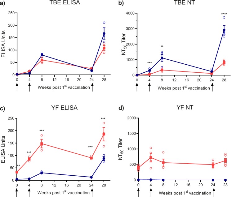
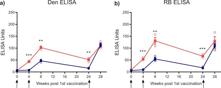
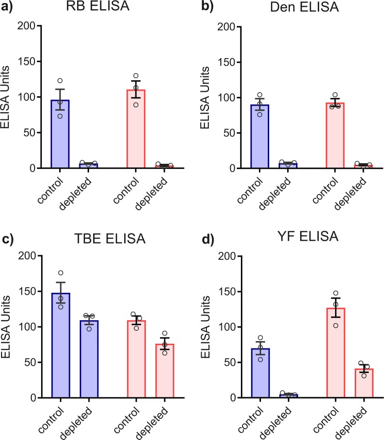
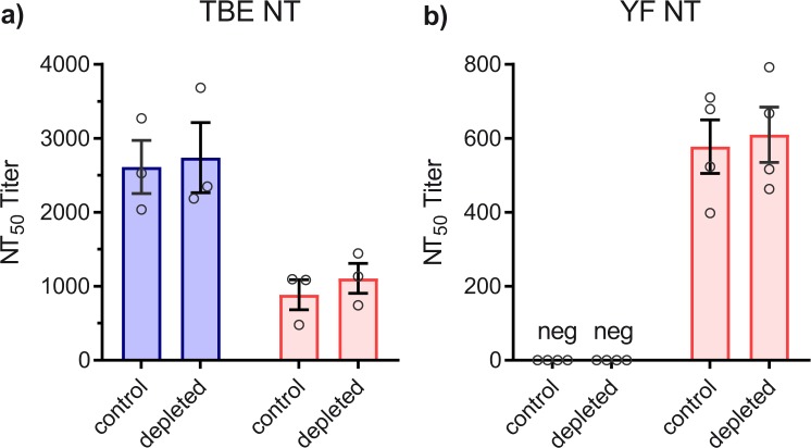
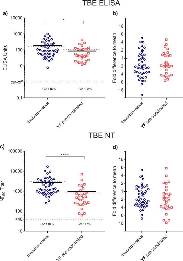
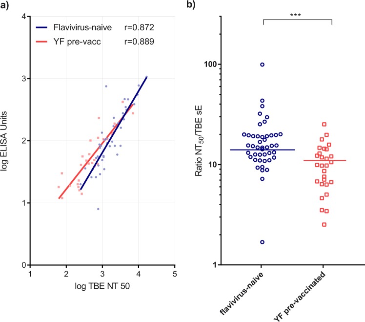
Flaviviruses have an increasing global impact as arthropod-transmitted human pathogens, exemplified by Zika, dengue, yellow fever (YF), West Nile, Japanese encephalitis, and tick-borne encephalitis (TBE) viruses. Since all flaviviruses are antigenically related, they are prone to phenomena of immunological memory ('original antigenic sin'), which can modulate immune responses in the course of sequential infections and/or vaccinations. In our study, we analyzed the influence of pre-existing YF vaccine-derived immunity on the antibody response to TBE vaccination. By comparing samples from YF pre-vaccinated and flavivirus-naive individuals, we show that YF immunity not only caused a significant impairment of the neutralizing antibody response to TBE vaccination but also a reduction of the specific TBE virus neutralizing activities (NT/ELISA-titer ratios). Our results point to a possible negative effect of pre-existing cross-reactive immunity on the outcome of flavivirus vaccination that may also pertain to other combinations of sequential flavivirus infections and/or vaccinations.
Keywords: Viral infection; Virology.
Conflict of interest statement
Competing interestsV.B., S.M., A.v.B., J.J., G.T., I.M., K.W., K.S. and F.X.H. declare no competing interests. U.K. declares to have the following competing interest: He has received travel grants and an unrestricted educational grants from Baxter Healthcare Inc. (Austria), which was the manufacturer of the TBE-vaccine used. He also declares that Baxter and its representatives had no influence on the planning, protocol and conduction of the study nor on the analysis and interpretation of the data.
Figures

Similar articles
-
Effect of previous heterologous flavivirus vaccinations on human antibody responses in tick-borne encephalitis and dengue virus infections.J Med Virol. 2023 Nov;95(11):e29245. doi: 10.1002/jmv.29245. J Med Virol. 2023. PMID: 38009693 Free PMC article.
-
Safety and immunogenicity of a purified inactivated Zika virus vaccine candidate in adults primed with a Japanese encephalitis virus or yellow fever virus vaccine in the USA: a phase 1, randomised, double-blind, placebo-controlled clinical trial.Lancet Infect Dis. 2023 Oct;23(10):1175-1185. doi: 10.1016/S1473-3099(23)00192-5. Epub 2023 Jun 27. Lancet Infect Dis. 2023. PMID: 37390836 Free PMC article. Clinical Trial.
-
Correlation between ELISA, hemagglutination inhibition, and neutralization tests after vaccination against tick-borne encephalitis.J Med Virol. 1996 Jan;48(1):102-7. doi: 10.1002/(SICI)1096-9071(199601)48:1<102::AID-JMV16>3.0.CO;2-I. J Med Virol. 1996. PMID: 8825718
-
Flaviviruses and their antigenic structure.J Clin Virol. 2012 Dec;55(4):289-95. doi: 10.1016/j.jcv.2012.08.024. Epub 2012 Sep 21. J Clin Virol. 2012. PMID: 22999801 Review.
-
Mucosal Vaccination: A Promising Alternative Against Flaviviruses.Front Cell Infect Microbiol. 2022 Jun 15;12:887729. doi: 10.3389/fcimb.2022.887729. eCollection 2022. Front Cell Infect Microbiol. 2022. PMID: 35782117 Free PMC article. Review.
Cited by
-
Genotype-specific features reduce the susceptibility of South American yellow fever virus strains to vaccine-induced antibodies.Cell Host Microbe. 2022 Feb 9;30(2):248-259.e6. doi: 10.1016/j.chom.2021.12.009. Epub 2022 Jan 7. Cell Host Microbe. 2022. PMID: 34998466 Free PMC article.
-
Distinct antibody repertoires against endemic human coronaviruses in children and adults.JCI Insight. 2021 Feb 22;6(4):e144499. doi: 10.1172/jci.insight.144499. JCI Insight. 2021. PMID: 33497357 Free PMC article.
-
Factors determining the outcomes of immune imprinting after repeated orthoflavivirus infections.Front Immunol. 2025 Jul 16;16:1560851. doi: 10.3389/fimmu.2025.1560851. eCollection 2025. Front Immunol. 2025. PMID: 40740782 Free PMC article. Review.
-
Schistosome and malaria exposure and urban-rural differences in vaccine responses in Uganda: a causal mediation analysis using data from three linked randomised controlled trials.Lancet Glob Health. 2024 Nov;12(11):e1860-e1870. doi: 10.1016/S2214-109X(24)00340-1. Lancet Glob Health. 2024. PMID: 39424574 Free PMC article. Clinical Trial.
-
Effect of previous heterologous flavivirus vaccinations on human antibody responses in tick-borne encephalitis and dengue virus infections.J Med Virol. 2023 Nov;95(11):e29245. doi: 10.1002/jmv.29245. J Med Virol. 2023. PMID: 38009693 Free PMC article.
References
-
- Bhatt Samir, Gething Peter W., Brady Oliver J., Messina Jane P., Farlow Andrew W., Moyes Catherine L., Drake John M., Brownstein John S., Hoen Anne G., Sankoh Osman, Myers Monica F., George Dylan B., Jaenisch Thomas, Wint G. R. William, Simmons Cameron P., Scott Thomas W., Farrar Jeremy J., Hay Simon I. The global distribution and burden of dengue. Nature. 2013;496(7446):504–507. doi: 10.1038/nature12060. - DOI - PMC - PubMed
Grants and funding
LinkOut - more resources
Full Text Sources
Research Materials
