. 2019 Jul 1;6(17):1900835.
doi: 10.1002/advs.201900835. eCollection 2019 Sep 4.

Cytomembrane-Mediated Transport of Metal Ions with Biological Specificity

Affiliations

Cytomembrane-Mediated Transport of Metal Ions with Biological Specificity

Ming-Kang Zhang et al. Adv Sci (Weinh). .

Abstract

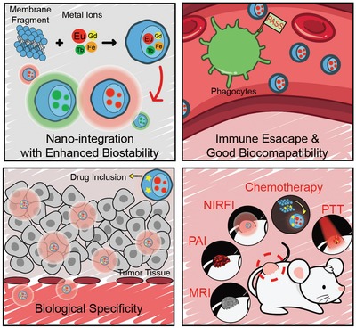
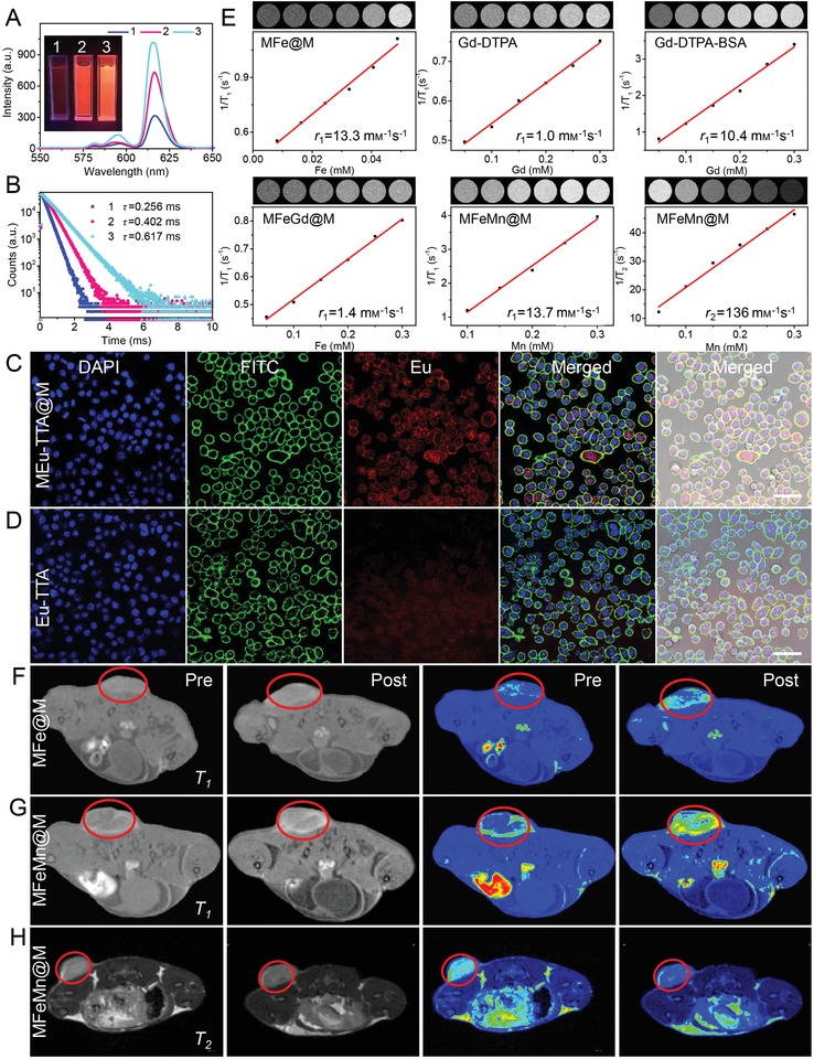
Metal ions are of significant importance in biomedical science. This study reports a new concept of cytomembrane-mediated biospecific transport of metal ions without using any other materials. For the first time, cytomembranes are exploited for two-step conjugation with metal ions to provide hybrid nanomaterials. The innate biofunction of cell membranes renders the hybrids with superior advantages over common vehicles for metal ions, including excellent biocompatibility, low immunogenic risk, and particularly specific biotargeting functionality. As a proof-of-concept demonstration, cancer cell membranes are used for in vivo delivery of various metal ions, including ruthenium, europium, iron, and manganese, providing a series of tumor-targeted nanohybrids capable of photothermal therapy/imaging, magnetic resonance imaging, photoacoustic imaging, and fluorescence imaging with improved performances. In addition, the special structure of the cell membrane allows easy accommodation of small-molecular agents within the nanohybrids for effective chemotherapy. This study provides a new class of metal-ion-included nanomaterials with versatile biofunctions and offers a novel solution to address the important challenge in the field of in vivo targeted delivery of metal ions.

Keywords: bioimaging; biotargeted transport; cell membrane; metal ions; tumor therapy.


Conflict of interest statement

The authors declare no conflict of interest.

Figures

Scheme 1
Scheme 1
Illustration of hybrid metal ion/cell membrane materials—biomimic transport of metal ion with biological specificity.
Figure 1
Figure 1
A) TEM images of MFe@M nanoparticles. B) UV–Vis spectra of CCMCs, FeCl3, and MFe@M. C) Photographs of FeCl3, CCMCs, and MFe@M before (C1) and after (C2) centrifugation and C3) photo images of supernatant of FeCl3, CCMCs, and MFe@M solutions after the centrifuging followed by the addition of Fehling reagent. D) Observation over the solution of RBC cells after the addition of FeCl3 and MFe@M. Concentrations of Fe3+ were 5, 10, 20, and 40 μg mL−1. E) Cellular internalization by macrophages after co‐incubation with MFe and MFe@M for 4 h. F) Cellular internalization assay of MFe@M‐4T1 in 4T1, B16, COS7, and HepG2 cells. The nucleus was stained blue with DAPI, the cell membrane was stained green with cell mask green, and MFe@M‐4T1 was stained red with DIR. Scale bar: 20 µm.
Figure 2
Figure 2
A) In vivo fluorescence images of 4T1‐tumor‐bearing mice and ex vivo fluorescence images of major organs after intravenously injected MFe@M‐4T1, MFe, CCMC‐4T1, and MFe@M‐CT26. B) Mean fluorescence intensity at tumor sites in the mice treated with MFe@M‐4T1, MFe, CCMC‐4T1, and MFe@M‐CT26. C) Mean fluorescence intensity of major organs in the mice treated with MFe@M‐4T1, MFe, CCMC‐4T1, and MFe@M‐CT26. (He, heart; Li, liver; Sp, spleen; Lu, lung; Ki, kidney; Tu, tumor). D) Photographs of tumors in different groups after treatment. E) Fluorescence images of tumor slices and photo images of major organs in the groups of MFe@M‐DOX and DOX. (Blue, DAPI; red, DOX) Scale bar: 50 µm. F) Mean fluorescence intensity of major organs in the groups of MFe@M‐DOX and DOX. G) Variation of relative tumor volume after different treatments.
Figure 3
Figure 3
A) Temperature curves of water, CCMCs, RuCl3, and MRu@M solutions upon the NIR irradiation at power density of (0.8 W cm−2). B) The temperature profile of MRu@M solution irradiated with 808‐nm laser, followed by natural cooling after the laser was turned off, C) determination of the system time constant using linear regression of the cooling profile shown in (B). D) Temperature curves and E) thermal photo images of the mice in groups of PBS, RuCl3, and MRu@M upon the NIR irradiation at power density of (0.8 W cm−2). F) Variation of average weight of the mice after photothermal therapy mediated with different samples. G) Variation of relative tumor volume after different treatments. H) Photographs of tumor in different groups after treatment. I) Images of PA signal of MRu@M nanoparticles at different concentration.
Figure 4
Figure 4
A) Photographs (insert) and fluorescence intensity of Eu‐TTA and MEu‐TTA@M. B) The decay curves of Eu‐TTA and MEu‐TTA@M. (1, Eu‐TTA in aqueous solution; 2, Eu‐TTA in ethanol; 3, MEu‐TTA@M in aqueous solution). C,D) Cellular internalization of MEu‐TTA@M (C) and Eu‐TTA (D) in 4T1 cells. Scale bar: 50 µm. E) T1‐MR images (insert) and plots of T1 relaxation rate (1/T1) in the solutions of MFe@M, Gd‐DTPA, Gd‐DTPA‐BSA, MFeGd@M, and MFeMn@M as well as T2‐MR images (insert) and plots of T2 relaxation rate (1/T2) in MFeMn@M solution. F) 2D axial T1‐weighted spin‐echo MR images and the corresponding color coding of MR images before (pre) and after (post) intravenous injection of MFe@M and G) MFeMn@M. H) 2D axial T2‐weighted spin‐echo MR images and corresponding color coding of MR images before (pre) and after (post) intravenous injection of MFeMn@M.

Similar articles

Cited by

References

    1. a) Thompson K. H., Orvig C., Science 2003, 300, 936; - PubMed
    2. b) Abrams M., Murrer B., Science 1993, 261, 725. - PubMed
    1. Guo Z., Sadler P. J., Angew. Chem., Int. Ed. 1999, 38, 1512. - PubMed
    1. a) Taylor‐Pashow K. M. L., Della Rocca J., Xie Z., Tran S., Lin W., J. Am. Chem. Soc. 2009, 131, 14261; - PMC - PubMed
    2. b) Horcajada P., Serre C., Maurin G., Ramsahye N. A., Balas F., Vallet‐Regí M., Sebban M., Taulelle F., Férey G., J. Am. Chem. Soc. 2008, 130, 6774; - PubMed
    3. c) Sun C.‐Y., Qin C., Wang C.‐G., Su Z.‐M., Wang S., Wang X.‐L., Yang G.‐S., Shao K.‐Z., Lan Y.‐Q., Wang E.‐B., Adv. Mater. 2011, 23, 5629. - PubMed
    1. a) Lauffer R. B., Chem. Rev. 1987, 87, 901;
    2. b) Qian X., Xu Z., Chem. Soc. Rev. 2015, 44, 4487; - PubMed
    3. c) Taylor K. M., Rieter W. J., Lin W., J. Am. Chem. Soc. 2008, 130, 14358; - PubMed
    4. d) Taylor K. M., Jin A., Lin W., Angew. Chem. 2008, 120, 7836. - PubMed
    1. a) Bornscheuer U. T., Kazlauskas R. J., Angew. Chem., Int. Ed. 2004, 43, 6032; - PubMed
    2. b) Steitz T. A., Steitz J. A., Proc. Natl. Acad. Sci. USA 1993, 90, 6498; - PMC - PubMed
    3. c) Khan M. T., Martell A. E., J. Am. Chem. Soc. 1967, 89, 4176; - PubMed
    4. d) Stadtman E. R., Free Radic. Biol. Med. 1990, 9, 315. - PubMed

LinkOut - more resources