

Quantitative ultrasound imaging monitoring progressive disuse osteopenia and mechanical stimulation mitigation in calcaneus region through a 90-day bed rest human study
- PMID: 31508307
- PMCID: PMC6718925
- DOI: 10.1016/j.jot.2018.11.004
Quantitative ultrasound imaging monitoring progressive disuse osteopenia and mechanical stimulation mitigation in calcaneus region through a 90-day bed rest human study
Abstract
Background: Osteoporosis parallels aging and functional mechanical unloading (e.g., space flight and bed rest), jeopardizing mineral density, microstructure, and integrity of bone and leading to an increased risk of fracture. A way to combat this deterioration is to harness the sensitivity of bone to mechanical signals.
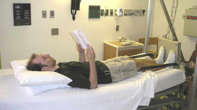
Objective: This study evaluates the longitudinal effect of a dynamic mechanical loading through the heel on human bone in vivo during 90-day bed rest, monitored by quantitative ultrasound (QUS) imaging and dual-energy X-ray absorptiometry (DXA) in localized regions of interests, i.e., calcaneus.
Methods: A total of 29 bed rest individuals were evaluated (11 control and 18 treatment) with a brief (10-minute) daily low-intensity (0.3g), high-frequency (30Hz) dynamic mechanical stimulation countermeasure through vibrational inhibition bone erosion (VIBE). Both QUS and DXA detected longitudinal bone density and quality changes.
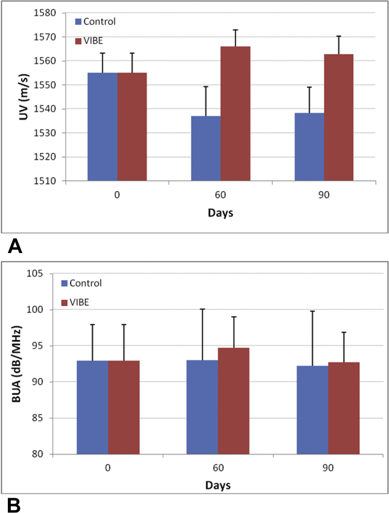
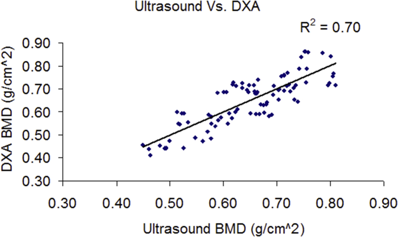
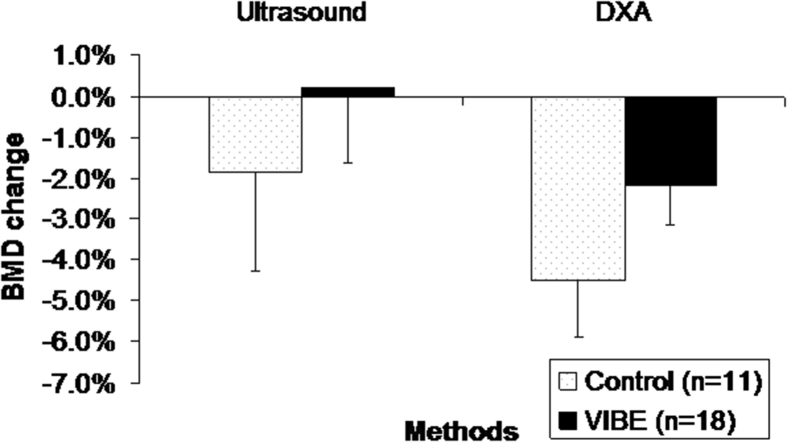
Results: Ultrasound velocity (UV) decreased in the control group and increased in the group treated with low-intensity loading. The UV increased by 1.9% and 1.6% at 60- and 90-day bed rest (p=0.01) in VIBE over control groups. A trend was found in broadband ultrasound attenuation (BUA), with a VIBE benefit of 1.8% at day 60 and 0.5% at day 90 in comparison with control (p=0.5). Bone mineral density (BMD) assessed by DXA decreased -4.50% for control individuals and -2.18% for VIBE individuals, showing a moderate effect of the mechanical intervention (p=0.19). Significant correlations between QUS and DXA were observed, with a combined BUA and UV vs. BMD: r2=0.70.
Conclusion: These results indicated that low-intensity, high-frequency loading has the potential to mitigate regional bone loss induced by long-term bed rest and that QUS imaging may be able to assess the subtle changes in bone alteration.
Translational potential of this article: Quantitative ultrasound has shown the efficacy of noninvasively assessing bone mass and structural properties in cadaver and isolated trabecular bone samples. While its ability in measuring in vivo bone quality and density is still unclear, a scanning confocal ultrasound imaging is developed and can perform an instant assessment for the subtle changes of such bone loss. This ultrasound imaging modality can potentially be used in the clinical assessment of bone mass. Moreover, physical stimulation has shown the ability to prevent bone loss induced by functional disuse and estrogen deficiency in animal models. However, its treatment capability is unclear. This study has shown that low-magnitude mechanical signals, introduced using low-intensity vibration (LIV), can mitigate regional bone loss caused by functional disuse. Thus localized mechanical treatment, and the quantitative ultrasound imaging have shown translational potential to noninvasively attenuate bone loss, and assess bone mass in the clinic, e.g., in an extreme condition such as long-term space mission, and long-term bedrest such as in case of spinal cord injury.
Keywords: Bed rest; Bone remodelling; Confocal acoustic navigation; Osteoporosis and osteopenia; Ultrasound imaging; Whole-body vibration.
Figures
References
-
- Arnaud S.B., Harper J.S., Navidi M. Mineral distribution in rat skeletons after exposure to a microgravity model. J Grav Physiol. 1995;2:115–116. - PubMed
-
- Arnaud S.B., Sherrard D.J., Maloney N., Whalen R.T., Fung P. Effects of 1-week head-down tilt bed rest on bone-formation and the calcium endocrine system. Aviat Space Environ Med. 1992;63:14–20. - PubMed
-
- Bauer D.C., Gluer C.C., Cauley J.A., Vogt T.M., Ensrud K.E., Genant H.K. Broadband ultrasound attenuation predicts fractures strongly and independently of densitometry in older women. A prospective study. Study of Osteoporotic Fractures Research Group. Arch Intern Med. 3-24-1997;157:629–634. - PubMed
-
- Belavy D.L., Miokovic T., Armbrecht G., Rittweger J., Felsenberg D. Resistive vibration exercise reduces lower limb muscle atrophy during 56-day bed-rest. J Musculoskelet Neuronal Interact. 2009;9:225–235. - PubMed
LinkOut - more resources
Full Text Sources
Research Materials
