

Discordance in diagnosis of osteoporosis by quantitative computed tomography and dual-energy X-ray absorptiometry in Chinese elderly men
- PMID: 31508308
- PMCID: PMC6718941
- DOI: 10.1016/j.jot.2018.11.003
Discordance in diagnosis of osteoporosis by quantitative computed tomography and dual-energy X-ray absorptiometry in Chinese elderly men
Abstract
Objective: The objective of this study was to investigate the diagnostic discordance of osteoporosis by quantitative computed tomography (QCT) and dual-energy X-ray absorptiometry (DXA) in Chinese elderly men.
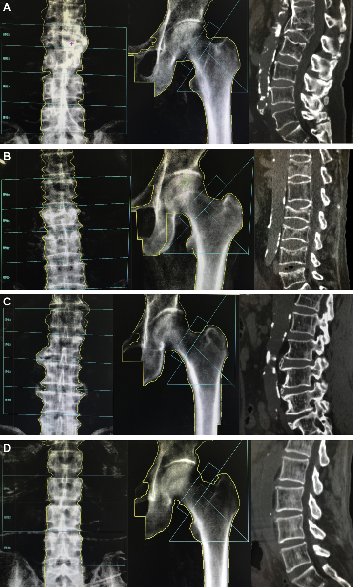
Methods: A total of 313 males older than 60 years, who underwent both spinal QCT and lumbar spine and hip DXA in our department, were included. The diagnostic criteria established by the World Health Organisation in 1994 were used for DXA to diagnose osteoporosis, and the criteria recommended by the International Society of Clinical Densitometry were used for QCT. The osteoporosis detection rate by the two techniques was calculated, and the difference was compared. The minor discordance was considered present when the different diagnostic classes between the two techniques were adjacent. Major discordance was present when the diagnosis by one technique was osteoporosis and the other was normal. The computed tomography images were reviewed by radiologists to assess whether vertebral fracture, aorta calcification or degeneration was present.
Results: In the 313 participants (mean age, 79.6 ± 7.2 years), the osteoporosis detection rate was 10.9% for DXA (lumbar spine and hip) and 45.1% for QCT, a significant difference (p < 0.001). The major discordance, minor discordance and concordance of diagnosis between the two techniques were seen in 8.3%, 50.8% and 40.9%, respectively. QCT detected osteoporosis better than DXA. The causes of this discordance were degeneration of spine, abdominal aorta calcification and vertebral fractures.
Conclusion: Our study demonstrated that discordance was common when using QCT and DXA to diagnose osteoporosis and that spinal degeneration, aorta calcification and fracture obscure the bone mineral density measurement of spine by DXA. QCT is a more sensitive method of choice to identify osteoporosis in elderly Chinese men.
The translational potential of this article: This study investigated the diagnostic discordance of osteoporosis by quantitative computed tomography (QCT) and dual-energy X-ray absorptiometry (DXA) in Chinese elderly men. The results demonstrated that QCT is a more sensitive method of choice to identify osteoporosis in elderly Chinese men. This work may help clinicians make an appropriate choice of technique for the accurate diagnosis of osteoporosis and identify the patients at high risk of osteoporosis who should be treated early to prevent fractures. This may influence the therapeutic plan and the overall prognosis of patients.
Keywords: Bone mineral density; Dual X-ray absorptiometry; Osteoporosis; Quantitative computed tomography.
Figures
Similar articles
-
Discordance in lumbar bone mineral density measurements by quantitative computed tomography and dual-energy X-ray absorptiometry in postmenopausal women: a prospective comparative study.Spine J. 2023 Feb;23(2):295-304. doi: 10.1016/j.spinee.2022.10.014. Epub 2022 Nov 4. Spine J. 2023. PMID: 36343911
-
Quantitative CT screening improved lumbar BMD evaluation in older patients compared to dual-energy X-ray absorptiometry.BMC Geriatr. 2023 Apr 17;23(1):231. doi: 10.1186/s12877-023-03963-6. BMC Geriatr. 2023. PMID: 37069511 Free PMC article.
-
Quantitative computed tomography has higher sensitivity detecting critical bone mineral density compared to dual-energy X-ray absorptiometry in postmenopausal women and elderly men with osteoporotic fractures: a real-life study.Arch Orthop Trauma Surg. 2024 Jan;144(1):179-188. doi: 10.1007/s00402-023-05070-y. Epub 2023 Oct 5. Arch Orthop Trauma Surg. 2024. PMID: 37796283
-
Role of dual-energy X-ray absorptiometry in the diagnosis and treatment of osteoporosis.J Clin Densitom. 2007 Jan-Mar;10(1):102-10. doi: 10.1016/j.jocd.2006.11.001. Epub 2006 Dec 27. J Clin Densitom. 2007. PMID: 17289532 Review.
-
X-ray-based quantitative osteoporosis imaging at the spine.Osteoporos Int. 2020 Feb;31(2):233-250. doi: 10.1007/s00198-019-05212-2. Epub 2019 Nov 14. Osteoporos Int. 2020. PMID: 31728606 Review.
Cited by
-
Chinese expert consensus on the diagnosis of osteoporosis by imaging and bone mineral density.Quant Imaging Med Surg. 2020 Oct;10(10):2066-2077. doi: 10.21037/qims-2020-16. Quant Imaging Med Surg. 2020. PMID: 33014734 Free PMC article.
-
MRI-based vertebral bone quality score as a novel bone status marker of patients with adolescent idiopathic scoliosis.Sci Rep. 2024 May 31;14(1):12518. doi: 10.1038/s41598-024-63426-9. Sci Rep. 2024. PMID: 38822099 Free PMC article.
-
Osteoporosis screening using QCT-based cutoff value of Hounsfield units in patients with degenerative lumbar diseases.Eur Spine J. 2024 Dec;33(12):4499-4503. doi: 10.1007/s00586-024-08491-4. Epub 2024 Sep 19. Eur Spine J. 2024. PMID: 39297897
-
The research progress on radionuclides in osteoporosis diagnosis and drug efficacy monitoring.Front Pharmacol. 2025 Apr 28;16:1594903. doi: 10.3389/fphar.2025.1594903. eCollection 2025. Front Pharmacol. 2025. PMID: 40356996 Free PMC article. Review.
-
A narrative review of the moderating effects and repercussion of exercise intervention on osteoporosis: ingenious involvement of gut microbiota and its metabolites.J Transl Med. 2022 Oct 27;20(1):490. doi: 10.1186/s12967-022-03700-4. J Transl Med. 2022. PMID: 36303163 Free PMC article. Review.
References
-
- Cummings S.R., Melton L.J. Epidemiology and outcomes of osteoporotic fractures. Lancet. 2002;359(9319):1761–1767. - PubMed
-
- Wade S.W., Strader C., Fitzpatrick L.A. Estimating prevalence of osteoporosis: examples from industrialized countries. Arch Osteoporos. 2014;9(1):182. - PubMed
-
- World Health Organization . Technical report series 843. WHO; Geneva: 1994. Assessment of fracture risk and its application to screening for postmenopausal osteoporosis. - PubMed
LinkOut - more resources
Full Text Sources
Research Materials
