

Radiomics-Based Machine Learning in Differentiation Between Glioblastoma and Metastatic Brain Tumors
- PMID: 31508366
- PMCID: PMC6714109
- DOI: 10.3389/fonc.2019.00806
Radiomics-Based Machine Learning in Differentiation Between Glioblastoma and Metastatic Brain Tumors
Abstract
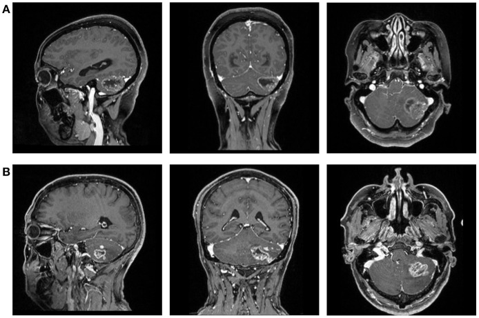
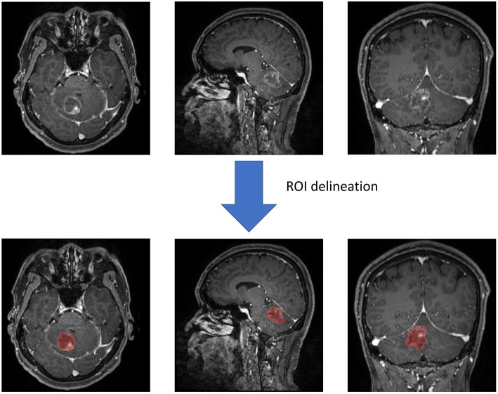
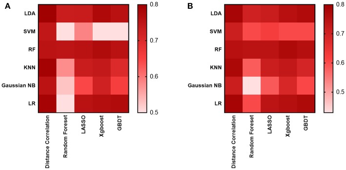
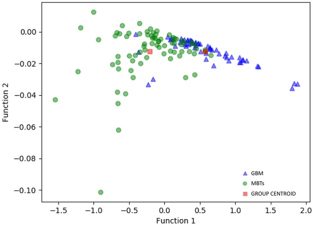
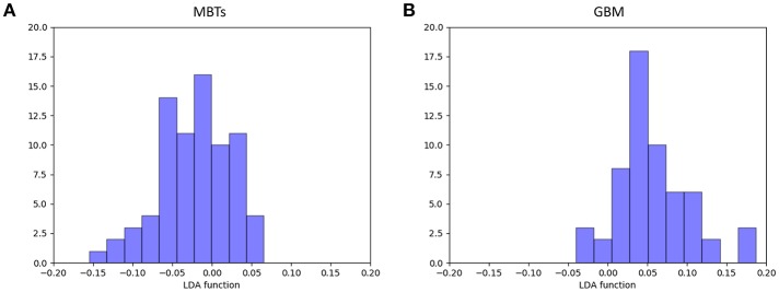
Purpose: To investigative the diagnostic performance of radiomics-based machine learning in differentiating glioblastomas (GBM) from metastatic brain tumors (MBTs). Method: The current study involved 134 patients diagnosed and treated in our institution between April 2014 and December 2018. Radiomics features were extracted from contrast-enhanced T1 weighted imaging (T1C). Thirty diagnostic models were built based on five selection methods and six classification algorithms. The sensitivity, specificity, accuracy, and area under curve (AUC) of each model were calculated, and based on these the optimal model was chosen. Result : Two models represented promising diagnostic performance with AUC of 0.80. The first model was a combination of Distance Correlation as the selection method and Linear Discriminant Analysis (LDA) as the classification algorithm. In the training group, the sensitivity, specificity, accuracy, and AUC were 0.75, 0.85, 0.80, and 0.80, respectively; and in the testing group, the sensitivity, specificity, accuracy, and AUC of the model were 0.69, 0.86, 0.78, and 0.80, respectively. The second model was the Distance Correlation as the selection method and logistic regression (LR) as the classification algorithm, with sensitivity, specificity, accuracy, and AUC of 0.75, 0.85, 0.80, 0.80 in the training group and 0.69, 0.86, 0.78, 0.80 in the testing group. Conclusion: Radiomic-based machine learning has potential to be utilized in differentiating GBM from MBTs.
Keywords: glioblastomas; machine learning; metastatic brain tumors; radiomics; texture analysis.
Figures
References
LinkOut - more resources
Full Text Sources
Miscellaneous