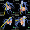
Can we delineate preoperatively the right and ventral margins of caudate lobe of the liver?
- PMID: 31508392
- PMCID: PMC6722294
- DOI: 10.4174/astr.2019.97.3.124
Can we delineate preoperatively the right and ventral margins of caudate lobe of the liver?
Abstract
Purpose: Complete removal of the caudate lobe, which is sometimes necessary, is accomplished via isolated caudate lobectomy or hepatectomy that includes the caudate lobe. It is impossible, however, to confirm the right and ventral margins of the caudate lobe by preoperative imaging. This study was undertaken to determine whether we could identify the right and ventral margins of the caudate lobe preoperatively using Synapse 3D visualization software.
Methods: Ninety-four preoperative 3-dimensional (3D) computed tomographic images (1-mm slices) of the liver from candidate donors were examined. The images of the caudate lobe were subjected to a counter-staining method according to Synapse 3D to delineate their dimensions. We first examined whether the right margin of the caudate lobe exceeded the plane formed by the root of the right hepatic vein (RHV) and the right side of the inferior vena cava (IVC). Second, we determined whether the ventral margin of the caudate lobe exceeded the plane formed by the root of the middle hepatic vein (MHV) and the root of the RHV.
Results: For the right margin, 17 cases (18%) exceeded the RHV-IVC plane by a mean of 10.2 mm (range, 2.4-27.2 mm). For the ventral margin, 28 cases (30%) exceeded the MHV-RHV plane by a mean of 17.4 mm (range, 1.2-49.1 mm).
Conclusion: Evaluating the anatomy of caudate lobe using Synapse 3D preoperatively could be helpful for more precise anatomical resection of the caudate lobe.
Keywords: Caudate lobe of liver; Liver anatomy; Preoperative; Three-dimensional imaging.
Conflict of interest statement
CONFLICTS OF INTEREST: No potential conflict of interest relevant to this article was reported.
Figures


Similar articles
-
Multi-detector row CT of relevant vascular anatomy of the surgical plane in split-liver transplantation.Radiology. 2003 Nov;229(2):401-7. doi: 10.1148/radiol.2292021437. Radiology. 2003. PMID: 14595144
-
Anatomical Boundary Between the Caudate Lobe of the Liver and Adjacent Segments Based on Three-Dimensional Analysis for Precise Resections.J Gastrointest Surg. 2018 Oct;22(10):1709-1714. doi: 10.1007/s11605-018-3819-5. Epub 2018 Jun 18. J Gastrointest Surg. 2018. PMID: 29916104
-
Location of the ventral margin of the paracaval portion of the caudate lobe of the human liver with special reference to the configuration of hepatic portal vein branches.Clin Anat. 2002 Nov;15(6):387-401. doi: 10.1002/ca.10055. Clin Anat. 2002. PMID: 12373729
-
Left trisectionectomy combined with resection of the right hepatic vein and inferior vena cava after right hepatic vein embolization for advanced intrahepatic cholangiocarcinoma.Surg Case Rep. 2019 Jun 18;5(1):98. doi: 10.1186/s40792-019-0655-0. Surg Case Rep. 2019. PMID: 31214903 Free PMC article.
-
The caudate lobe of the liver: implications of embryology and anatomy for surgery.Surg Oncol Clin N Am. 2002 Oct;11(4):835-48. doi: 10.1016/s1055-3207(02)00035-2. Surg Oncol Clin N Am. 2002. PMID: 12607574 Review.
Cited by
-
Resection of the paracaval portion of the caudate lobe of the liver through complete right approach: advantages of a laparoscopic perspective.Updates Surg. 2025 Apr;77(2):455-458. doi: 10.1007/s13304-025-02134-z. Epub 2025 Mar 3. Updates Surg. 2025. PMID: 40032804
-
ASO Author Reflections: Real Anatomical Right Hepatectomy Preserving the Caudate Lobe: Advancing Precision in Liver Surgery.Ann Surg Oncol. 2025 Apr;32(4):2487-2488. doi: 10.1245/s10434-025-16887-9. Epub 2025 Jan 17. Ann Surg Oncol. 2025. PMID: 39821550 No abstract available.
-
A practical study of the hepatic vascular system anatomy of the caudate lobe.Quant Imaging Med Surg. 2021 Apr;11(4):1313-1321. doi: 10.21037/qims-20-780. Quant Imaging Med Surg. 2021. PMID: 33816170 Free PMC article.
-
Robotic Real Anatomical Right Hepatectomy Preserving the Caudate Lobe: Separate Dissection of the Right Anterior and Posterior Glissonean Pedicles, Combined with the Use of ICG Fluorescent Imaging (with Video).Ann Surg Oncol. 2025 Apr;32(4):2467-2471. doi: 10.1245/s10434-024-16761-0. Epub 2024 Dec 27. Ann Surg Oncol. 2025. PMID: 39730961
References
-
- Mizumoto R, Kawarada Y, Suzuki H. Surgical treatment of hilar carcinoma of the bile duct. Surg Gynecol Obstet. 1986;162:153–158. - PubMed
-
- Baton O, Azoulay D, Adam DV, Castaing D. Major hepatectomy for hilar cholangiocarcinoma type 3 and 4: prognostic factors and longterm outcomes. J Am Coll Surg. 2007;204:250–260. - PubMed
-
- Couinaud C. Liver lobes and segments: notes on the anatomical architecture and surgery of the liver. Presse Med. 1954;62:709–712. - PubMed
-
- Kumon M. Anatomy of the caudate lobe with special reference to portal vein and bile duct. Acta Hepatol Jpn. 1985;26:1193–1199.
LinkOut - more resources
Full Text Sources

