

Influence of Fixation and Permeabilization on the Mass Density of Single Cells: A Surface Plasmon Resonance Imaging Study
- PMID: 31508410
- PMCID: PMC6716545
- DOI: 10.3389/fchem.2019.00588
Influence of Fixation and Permeabilization on the Mass Density of Single Cells: A Surface Plasmon Resonance Imaging Study
Abstract
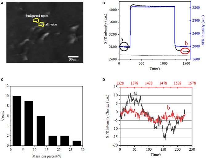
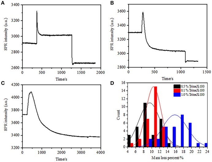
Fixation and permeabilization of cells and tissues are essential processes in biological techniques like immunofluorescence and immunohistochemistry for cell biology studies. In typical procedures, the biological samples are treated by paraformaldehyde and Triton X-100 to achieve cellular fixation and permeabilization, respectively, prior to the incubation with specific antibodies. While it is well-known that the integrity of cell membrane has been broken during these processes, quantitative studies on the loss of cellular mass density and the enhancement of molecular accessibility at single cell level are still rare. In this study, we employed the surface plasmon resonance (SPR) imaging technique to monitor the mass density change of single cells during sequential fixation and permeabilization processes. We further utilize the osmotic responses of single cells to sugar molecules as an indicator to evaluate the integrity of cell membranes. It was found that, while fixation initially destructed the integrity of cell membranes and increased the permeability of intra- and extra-cellular molecules, it was permeabilization process that substantially induced significant loss in cellular mass density.
Keywords: fixation; immunofluorescence; osmotic pressure; permeabilization; surface plasmon resonance imaging.
Figures


Similar articles
-
Sequential paraformaldehyde and methanol fixation for simultaneous flow cytometric analysis of DNA, cell surface proteins, and intracellular proteins.Cytometry. 1992;13(4):432-44. doi: 10.1002/cyto.990130414. Cytometry. 1992. PMID: 1382010
-
Rapid single-step method for flow cytometric detection of surface and intracellular antigens using whole blood.Cytometry. 1996 Sep 1;25(1):58-70. doi: 10.1002/(SICI)1097-0320(19960901)25:1<58::AID-CYTO7>3.0.CO;2-A. Cytometry. 1996. PMID: 8875055
-
Redistribution and differential extraction of soluble proteins in permeabilized cultured cells. Implications for immunofluorescence microscopy.J Cell Sci. 1992 Apr;101 ( Pt 4):731-43. doi: 10.1242/jcs.101.4.731. J Cell Sci. 1992. PMID: 1527176
-
Membrane Repair Mechanisms against Permeabilization by Pore-Forming Toxins.Toxins (Basel). 2018 Jun 9;10(6):234. doi: 10.3390/toxins10060234. Toxins (Basel). 2018. PMID: 29890730 Free PMC article. Review.
-
Surface plasmon resonance: a label-free tool for cellular analysis.Nanomedicine (Lond). 2015;10(11):1833-46. doi: 10.2217/nnm.15.31. Nanomedicine (Lond). 2015. PMID: 26080702 Review.
Cited by
-
Optimization of Neurite Tracing and Further Characterization of Human Monocyte-Derived-Neuronal-like Cells.Brain Sci. 2021 Oct 20;11(11):1372. doi: 10.3390/brainsci11111372. Brain Sci. 2021. PMID: 34827371 Free PMC article.
-
Impact of Triton X-100 on Notch 1 Surface Receptor Immunofluorescence: A Cautionary Study.Discoveries (Craiova). 2025 Mar 31;13(1):e206. doi: 10.15190/d.2025.5. eCollection 2025 Jan-Mar. Discoveries (Craiova). 2025. PMID: 40351502 Free PMC article.
-
A Mammalian Conserved Circular RNA CircLARP1B Regulates Hepatocellular Carcinoma Metastasis and Lipid Metabolism.Adv Sci (Weinh). 2024 Jan;11(2):e2305902. doi: 10.1002/advs.202305902. Epub 2023 Nov 12. Adv Sci (Weinh). 2024. PMID: 37953462 Free PMC article.
-
Subcellular proteomics.Nat Rev Methods Primers. 2021;1:32. doi: 10.1038/s43586-021-00029-y. Epub 2021 Apr 29. Nat Rev Methods Primers. 2021. PMID: 34549195 Free PMC article.
-
Bypassing the Need for Cell Permeabilization: Nanobody CDR3 Peptide Improves Binding on Living Bacteria.Bioconjug Chem. 2023 Jul 19;34(7):1234-1243. doi: 10.1021/acs.bioconjchem.3c00116. Epub 2023 Jul 7. Bioconjug Chem. 2023. PMID: 37418494 Free PMC article.
References
LinkOut - more resources
Full Text Sources
Other Literature Sources