

A Direct Comparison of Biplanar Videoradiography and Optical Motion Capture for Foot and Ankle Kinematics
- PMID: 31508415
- PMCID: PMC6716496
- DOI: 10.3389/fbioe.2019.00199
A Direct Comparison of Biplanar Videoradiography and Optical Motion Capture for Foot and Ankle Kinematics
Abstract
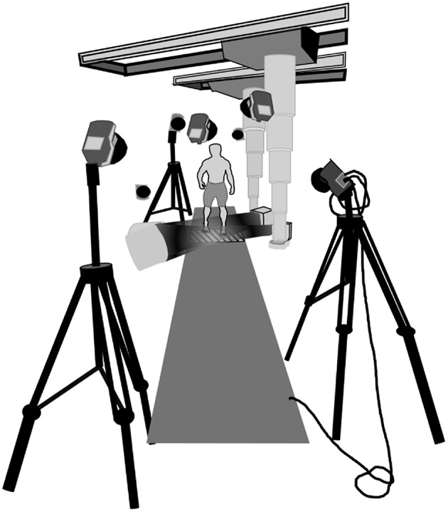
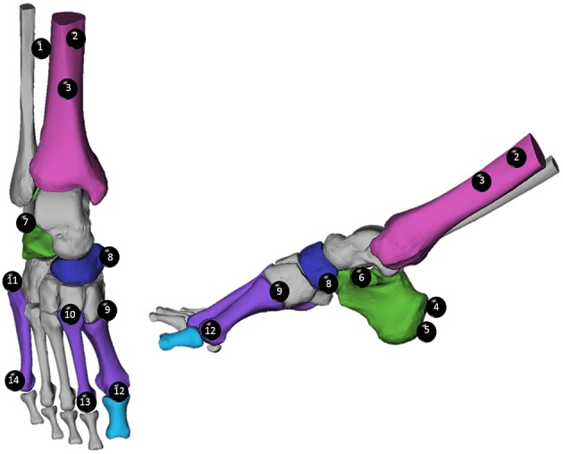
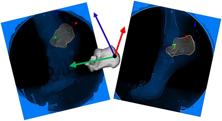
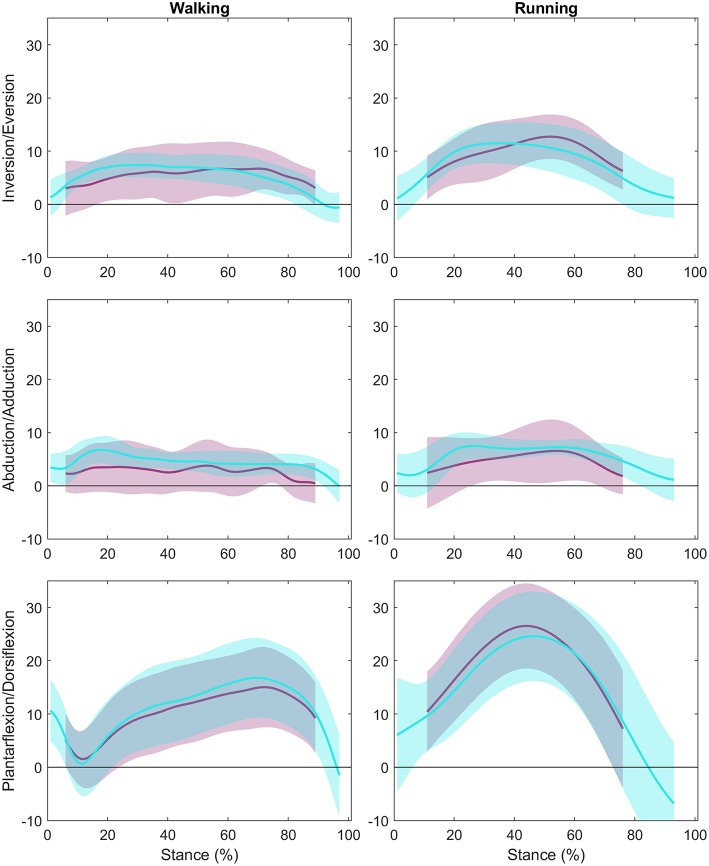
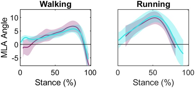
Measuring motion of the human foot presents a unique challenge due to the large number of closely packed bones with congruent articulating surfaces. Optical motion capture (OMC) and multi-segment models can be used to infer foot motion, but might be affected by soft tissue artifact (STA). Biplanar videoradiography (BVR) is a relatively new tool that allows direct, non-invasive measurement of bone motion using high-speed, dynamic x-ray images to track individual bones. It is unknown whether OMC and BVR can be used interchangeably to analyse multi-segment foot motion. Therefore, the aim of this study was to determine the agreement in kinematic measures of dynamic activities. Nine healthy participants performed three walking and three running trials while BVR was recorded with synchronous OMC. Bone position and orientation was determined through manual scientific-rotoscoping. The OMC and BVR kinematics were co-registered to the same coordinate system, and BVR tracking was used to create virtual markers for comparison to OMC during dynamic trials. Root mean square (RMS) differences in marker positions and joint angles as well as a linear fit method (LFM) was used to compare the outputs of both methods. When comparing BVR and OMC, sagittal plane angles were in good agreement (ankle: R2 = 0.947, 0.939; Medial Longitudinal Arch (MLA) Angle: R2 = 0.713, 0.703, walking and running, respectively). When examining the ankle, there was a moderate agreement between the systems in the frontal plane (R2 = 0.322, 0.452, walking and running, respectively), with a weak to moderate correlation for the transverse plane (R2 = 0.178, 0.326, walking and running, respectively). However, root mean squared error (RMSE) showed angular errors ranging from 1.06 to 8.31° across the planes (frontal: 3.57°, 3.67°, transverse: 4.28°, 4.70°, sagittal: 2.45°, 2.67°, walking and running, respectively). Root mean square (RMS) differences between OMC and BVR marker trajectories were task dependent with the largest differences in the shank (6.0 ± 2.01 mm) for running, and metatarsals (3.97 ± 0.81 mm) for walking. Based on the results, we suggest BVR and OMC provide comparable solutions to foot motion in the sagittal plane, however, interpretations of out-of-plane movement should be made carefully.
Keywords: ankle; biplanar videoradiography; foot; kinematics; motion analysis.
Figures
References
-
- Brainerd E. L., Baier D. B., Gatesy S. M., Hedrick T. L., Metzger K. A., Gilbert S. L., et al. . (2010). X-ray reconstruction of moving morphology (XROMM): precision, accuracy and applications in comparative biomechanics research. J. Exp. Zool. A Ecol. Genet. Physiol. 313, 262–279. 10.1002/jez.589 - DOI - PubMed
LinkOut - more resources
Full Text Sources