

Body motion detection and correction in cardiac PET: Phantom and human studies
- PMID: 31508827
- PMCID: PMC6842053
- DOI: 10.1002/mp.13815
Body motion detection and correction in cardiac PET: Phantom and human studies
Abstract
Purpose: Patient body motion during a cardiac positron emission tomography (PET) scan can severely degrade image quality. We propose and evaluate a novel method to detect, estimate, and correct body motion in cardiac PET.
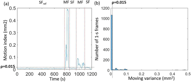
Methods: Our method consists of three key components: motion detection, motion estimation, and motion-compensated image reconstruction. For motion detection, we first divide PET list-mode data into 1-s bins and compute the center of mass (COM) of the coincidences' distribution in each bin. We then compute the covariance matrix within a 25-s sliding window over the COM signals inside the window. The sum of the eigenvalues of the covariance matrix is used to separate the list-mode data into "static" (i.e., body motion free) and "moving" (i.e. contaminated by body motion) frames. Each moving frame is further divided into a number of evenly spaced sub-frames (referred to as "sub-moving" frames), in which motion is assumed to be negligible. For motion estimation, we first reconstruct the data in each static and sub-moving frame using a rapid back-projection technique. We then select the longest static frame as the reference frame and estimate elastic motion transformations to the reference frame from all other static and sub-moving frames using nonrigid registration. For motion-compensated image reconstruction, we reconstruct all the list-mode data into a single image volume in the reference frame by incorporating the estimated motion transformations in the PET system matrix. We evaluated the performance of our approach in both phantom and human studies.
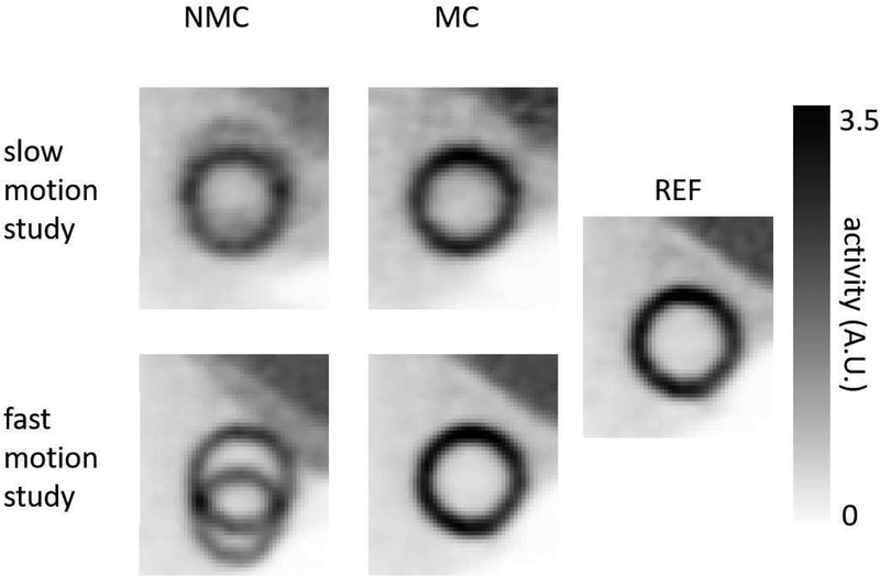
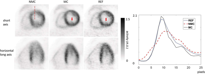
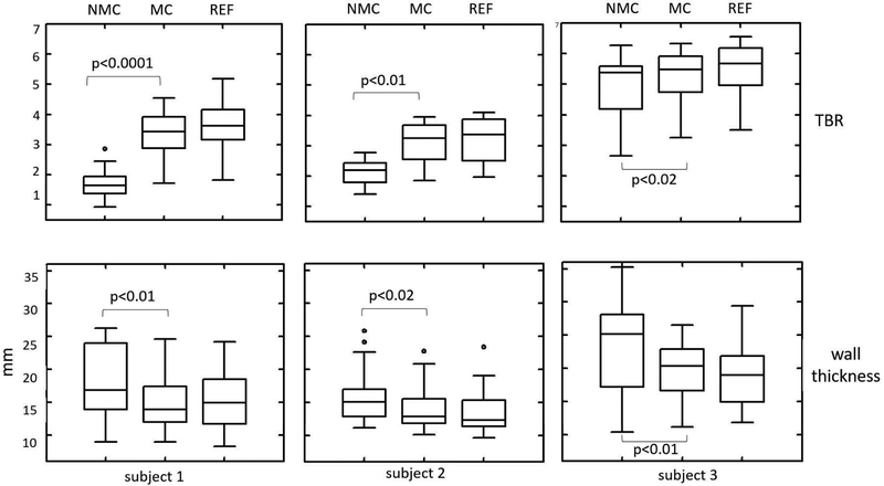
Results: Visually, the motion-corrected (MC) PET images obtained using the proposed method have better quality and fewer motion artifacts than the images reconstructed without motion correction (NMC). Quantitative analysis indicates that MC yields higher myocardium to blood pool concentration ratios. MC also yields sharper myocardium than NMC.
Conclusions: The proposed body motion correction method improves image quality of cardiac PET.
Keywords: body motion; bulk motion; cardiac PET; image reconstruction; motion correction; motion detection; motion estimation.
© 2019 American Association of Physicists in Medicine.
Conflict of interest statement
CONFLICT OF INTEREST
The authors have no conflicts to disclose.
Figures

Similar articles
-
MR-based cardiac and respiratory motion correction of PET: application to static and dynamic cardiac 18F-FDG imaging.Phys Med Biol. 2019 Oct 4;64(19):195009. doi: 10.1088/1361-6560/ab39c2. Phys Med Biol. 2019. PMID: 31394518 Free PMC article.
-
Patient motion effects on the quantification of regional myocardial blood flow with dynamic PET imaging.Med Phys. 2016 Apr;43(4):1829. doi: 10.1118/1.4943565. Med Phys. 2016. PMID: 27036580
-
Improved frame-based estimation of head motion in PET brain imaging.Med Phys. 2016 May;43(5):2443. doi: 10.1118/1.4946814. Med Phys. 2016. PMID: 27147355 Free PMC article.
-
Magnetic resonance-based motion correction for positron emission tomography imaging.Semin Nucl Med. 2013 Jan;43(1):60-7. doi: 10.1053/j.semnuclmed.2012.08.007. Semin Nucl Med. 2013. PMID: 23178089 Free PMC article. Review.
-
MR-Based Cardiac and Respiratory Motion-Compensation Techniques for PET-MR Imaging.PET Clin. 2016 Apr;11(2):179-91. doi: 10.1016/j.cpet.2015.09.004. Epub 2016 Jan 26. PET Clin. 2016. PMID: 26952730 Review.
Cited by
-
Improvement of motion artifacts using dynamic whole-body 18F-FDG PET/CT imaging.Jpn J Radiol. 2024 Apr;42(4):374-381. doi: 10.1007/s11604-023-01513-z. Epub 2023 Dec 14. Jpn J Radiol. 2024. PMID: 38093138 Free PMC article.
-
Inter-pass motion correction for whole-body dynamic PET and parametric imaging.IEEE Trans Radiat Plasma Med Sci. 2023 Apr;7(4):344-353. doi: 10.1109/trpms.2022.3227576. Epub 2022 Dec 8. IEEE Trans Radiat Plasma Med Sci. 2023. PMID: 37842204 Free PMC article.
-
The value of dynamic FDG PET/CT in the differential diagnosis of lung cancer and predicting EGFR mutations.BMC Pulm Med. 2024 May 10;24(1):227. doi: 10.1186/s12890-024-02997-9. BMC Pulm Med. 2024. PMID: 38730287 Free PMC article.
-
Impact of motion correction on [18F]-MK6240 tau PET imaging.Phys Med Biol. 2023 May 15;68(10):10.1088/1361-6560/acd161. doi: 10.1088/1361-6560/acd161. Phys Med Biol. 2023. PMID: 37116511 Free PMC article.
-
Synergistic motion compensation strategies for positron emission tomography when acquired simultaneously with magnetic resonance imaging.Philos Trans A Math Phys Eng Sci. 2021 Aug 23;379(2204):20200207. doi: 10.1098/rsta.2020.0207. Epub 2021 Jul 5. Philos Trans A Math Phys Eng Sci. 2021. PMID: 34218675 Free PMC article. Review.
References
-
- Gillman A, Smith J, Thomas P, Rose S, and Dowson N, PET motion correction in context of integrated PET/MR: Current techniques, limitations, and future projections: Current, Med. Phys 44(12), 430–445 (2017). - PubMed
-
- Carles M, Bach T, Torres-Espallardo I, Baltas D, Nestle U, and Martí-Bonmatí L, Significance of the impact of motion compensation on the variability of PET image features, Phys. Med. Biol 63(6), 065013 (2018). - PubMed
MeSH terms
Substances
Grants and funding
LinkOut - more resources
Full Text Sources
Other Literature Sources
