Case Reports
. 2019 Oct;6(10):2104-2109.
doi: 10.1002/acn3.50858. Epub 2019 Sep 11.

Responsive neurostimulation targeting anterior thalamic nucleus in generalized epilepsy

Affiliations
Case Reports

Responsive neurostimulation targeting anterior thalamic nucleus in generalized epilepsy

Aline Herlopian et al. Ann Clin Transl Neurol. 2019 Oct.

Abstract

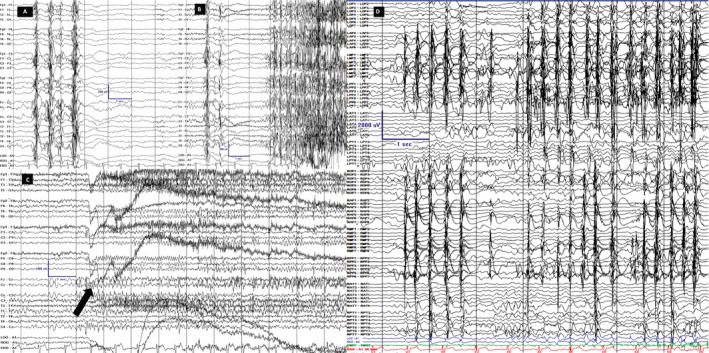
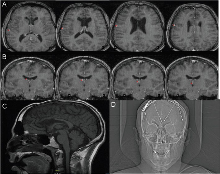
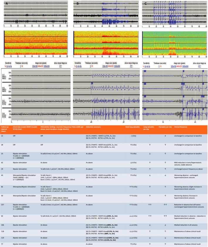
Responsive neurostimulation (RNS) has emerged as an adjunctive treatment modality for patients with intractable focal epilepsy who are not surgical candidates or have more than one ictal onset focus. We report a 34-year-old patient with intractable, childhood-onset, genetic generalized epilepsy (GGE) with tonic, atonic, myoclonic and absence seizures treated with RNS. Strip electrodes over the right posterior frontal cortex and depth electrodes placed in the right anterior nucleus were used for event detection and responsive stimulation. Two-year follow-up revealed 90-95% clinical seizure reduction. This case suggests that refractory GGE may be effectively treated with RNS targeting thalamocortical networks.


Conflict of interest statement

Dr Andrew Cole received coverage for travel expenses but no honorarium for a Neuropace advisory meeting. Dr Aline Herlopian received travel funding during fellowship to attend a training conference on responsive neurostimulation sponsored by Neuropace. None of the other authors has any conflict of interest to disclose pertinent to this article.

Figures

Figure 1
Figure 1
Scalp and invasive EEG recordings. LFF 1Hz; HFF 70Hz, Notch OFF; Sensitivity 7 uV/mm Timebase 30mm/sec. Scale is displayed in all images; y‐axis is amplitude in uV and x‐axis is time in seconds. Scalp (A–C) and intracranial (D) recordings are on longitudinal bipolar montage along with coronal ring on scalp. A. Interictal background displayed generalized PSSW (polyspike, spike and slow wave) discharges without hemispheric predominance B. Tonic seizure. The first unequivocal ictal electrographic change occurred as burst of generalized PSSW followed by electro‐decrement and generalized FFA (faster frequency activity). The first ictal behavior occurred 2‐3 seconds after the first EEG change. C. Hypermotoric seizure. The first unequivocal electrographic change occurred as generalized, centrally predominant, medium amplitude FFA (arrow) and clinically evidenced by eyelid myoclonia followed by myoclonus of legs and flailing of left hand. D. Typical brief hypermotoric and myoclonic seizure evidenced at onset by synchronous periodic discharges from the RPF (right posterior frontal)/ RMF (right middle frontal)/ RAF (right anterior frontal)/ ROF (right orbitofrontal)/ LMF (left middle frontal)/ LAF (left anterior frontal)/ LPF (left posterior frontal) highest in amplitude on the lateral contacts.
Figure 2
Figure 2
(A–B) 3D reconstruction of the T1‐MRI brain sequences after RNS implantation (A) Axial T1‐sequences revealing the annotations of the strip placed on the right posterior frontal cortex (rPFC) contact with the first image revealing first contact while the last sequence shows the last contact on the strip # 4. (B) Coronal T1‐sequences revealing the depth electrode to the right anterior thalamic nucleus (rATN) with the first image revealing contact # 4 that is the most lateral contact and the last image is contact # 1 that is the deepest contact (C) MRI T1‐sagittal view displaying anterior corpus callosotomy changes. (D) An X‐ray of head displaying the implantation of RNS device with 2‐depth electrodes each with four contacts targeting the ATN and 2‐strips at the PFC. Only the rATN depth and rPFC strip were connected to the RNS device.
Figure 3
Figure 3
Data from the responsive neurostimulation (RNS). (1) (A–C) Electrocorticography (ECOG) from the RNS device and (2) table depicting various stimulation/detection parameters with corresponding ECOG and clinical response. (A–C) Channel (Ch) 1 and 2 are the deepest contacts of the depth electrode targeting the right anterior thalamic nucleus (rATN). Channel (Ch) 3 and 4 are the most posterior contacts on the right posterior frontal cortex (rPFC) strip. A1:Prestimulation baseline background recording from both rATN and rPFC. A2 is a magnification of A1. A3 represents the spectral analysis during prestimulation interictal phase. B1:Prestimulation baseline ictal recording from both rATN and rPFC. The seizures start synchronously from both rATN and rPFC. B2 is a magnified ECOG of the ictal activity. B3. Represents the spectral analysis during prestimulation ictal phase. C1. Stimulation parameters were turned on delivering burst therapy to both rATN and rPFC contacts to abort the seizure. Stimulation was delivered in bipolar mode. C2 is a magnified display of the treatment bursts delivered. Note the ictal onset occuring in rPFC 0.5 seconds prior to recruitment of rATN. C3. Represents the spectral analysis during ictal phase stimulation. Table Legend: TX: Treatment; ↓:mild decrease; ↓↓: marked decreased; ↑:mild increase; ↑↑: marked increase.

Similar articles

Cited by

References

    1. Morrell MJ. Responsive cortical stimulation for the treatment of medically intractable partial epilepsy. Neurology 2011;77:1295–1304. - PubMed
    1. Bergey GK, Morrell MJ, Mizrahi EM, et al. Long‐term treatment with responsive brain stimulation in adults with refractory partial seizures. Neurology 2015;84:810–817. - PMC - PubMed
    1. Meeren H, van Luijtelaar G, Lopes da Silva F, Coenen A. Evolving concepts on the pathophysiology of absence seizures: the cortical focus theory. Arch Neurol 2005;62:371–376. - PubMed
    1. Pinault D, O’Brien TJ. Cellular and network mechanisms of genetically‐determined absence seizures. Thalamus Relat Syst 2005;3:181–203. - PMC - PubMed
    1. Mirski MA, Rossell LA, Terry JB, Fisher RS. Anticonvulsant effect of anterior thalamic high frequency electrical stimulation in the rat. Epilepsy Res 1997;28:89–100. - PubMed

Publication types

LinkOut - more resources