Parathyromatosis: a very rare cause of recurrent primary hyperparathyroidism - case report and review of the literature
- PMID: 31509000
- PMCID: PMC6818056
- DOI: 10.1308/rcsann.2019.0105
Parathyromatosis: a very rare cause of recurrent primary hyperparathyroidism - case report and review of the literature
Abstract
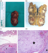
Parathyromatosis is a rare entity and usually appears as a consequence of the seeding on previous parathyroid surgery which was applied for the secondary hyperparathyroidism. A 63-year-old woman presented with a history of subtotal thyroidectomy 20 years ago and parathyroidectomy due to primary hyperparathyroidism (PHPT) four years ago. Imaging methods revealed multiple parathyromatosis foci on subcutaneous tissue of the neck. En-bloc resection was performed and pathological examination confirmed the diagnosis of parathyromatosis. After an uneventful 10 months, biochemical and radiological tests revealed recurrence on bilateral thyroid lodges. En-bloc resection was performed. The patient has remained well for 24 months after the second operation and has been followed-up with normal parathormone and serum calcium values. To the best of our knowledge, this report describes the twenty-first case of parathyromatosis in PHPT setting in the literature. It should be kept in mind that parathyromatosis may recur at different sites in the neck even in patients with PHPT.
Keywords: Parathyroidectomy; Parathyromatosis; Primary hyperparathyroidism; Recurrent parathyromatosis.
Figures


Similar articles
-
Parathyromatosis: a cause for recurrent hyperparathyroidism.Endocr Pract. 2001 May-Jun;7(3):189-92. doi: 10.4158/EP.7.3.189. Endocr Pract. 2001. PMID: 11421566 Review.
-
Recurrent primary hyperparathyroidism due to Type 1 parathyromatosis.Endocrine. 2017 Feb;55(2):643-650. doi: 10.1007/s12020-016-1139-7. Epub 2016 Oct 14. Endocrine. 2017. PMID: 27743301
-
Recurrent renal hyperparathyroidism caused by parathyromatosis.World J Surg. 2007 Feb;31(2):299-305. doi: 10.1007/s00268-006-0391-z. World J Surg. 2007. PMID: 17219279
-
Recurrent hyperparathyroidism secondary to parathyromatosis: clinical and imaging findings.J Ultrasound Med. 2007 Jun;26(6):847-51. doi: 10.7863/jum.2007.26.6.847. J Ultrasound Med. 2007. PMID: 17526617
-
Recurrent hyperparathyroidism caused by benign neoplastic seeding: two cases of parathyromatosis and a review of the literature.Acta Chir Belg. 2013 May-Jun;113(3):228-32. doi: 10.1080/00015458.2013.11680918. Acta Chir Belg. 2013. PMID: 24941723 Review.
Cited by
-
Retrosternal parathyromatosis in a patient with prior total parathyroidectomy.J Surg Case Rep. 2023 Jun 5;2023(6):rjad256. doi: 10.1093/jscr/rjad256. eCollection 2023 Jun. J Surg Case Rep. 2023. PMID: 37293335 Free PMC article.
-
Recurrent secondary hyperparathyroidism after parathyroidectomy due to anterior mediastinum ectopic parathyroid glands in a peritoneal dialysis patient-a case report and literature review.Front Med (Lausanne). 2025 Jun 16;12:1564135. doi: 10.3389/fmed.2025.1564135. eCollection 2025. Front Med (Lausanne). 2025. PMID: 40589961 Free PMC article.
-
A case of mediastinal hyperparathyromatosis.J Surg Case Rep. 2024 Jan 18;2024(1):rjad735. doi: 10.1093/jscr/rjad735. eCollection 2024 Jan. J Surg Case Rep. 2024. PMID: 38250132 Free PMC article.
-
Hypercalcemia during pregnancy: management and outcomes for mother and child.Endocrine. 2021 Mar;71(3):604-610. doi: 10.1007/s12020-021-02615-2. Epub 2021 Feb 5. Endocrine. 2021. PMID: 33544354 Free PMC article.
-
European Expert Consensus on Practical Management of Specific Aspects of Parathyroid Disorders in Adults and in Pregnancy: Recommendations of the ESE Educational Program of Parathyroid Disorders.Eur J Endocrinol. 2022 Jan 13;186(2):R33-R63. doi: 10.1530/EJE-21-1044. Print 2022 Feb 1. Eur J Endocrinol. 2022. PMID: 34863037 Free PMC article.
References
-
- Fernandez-Ranvier GG, Khanafshar E, Jensen K et al. . Parathyroid carcinoma, atypical parathyroid adenoma, or parathyromatosis? Cancer 2007; : 255–264. - PubMed
-
- Baloch ZW, Fraker D, LiVolsi VA. Parathyromatosis as cause of recurrent secondary hyperparathyroidism: a cytologic diagnosis. Diagn Cytopathol 2001; : 403–405. - PubMed
-
- Reddick RL, Costa JC, Marx SJ. Parathyroid hyperplasia and parathyromatosis. Lancet 1977; (i): 549. - PubMed
-
- Rattner DW, Marrone GC, Kasdon K et al. . Recurrent hyperparathyroidism due to implantation of parathyroid tissue. Am J Surg 1985; : 745–748. - PubMed
-
- Akerstrom G, Rudberg C, Grimelius K et al. . Recurrent hyperparathyroidism due to peroperative seeding of neoplastic or hyperplastic parathyroid tissue: case report. Acta Chir Scand 1988; : 549–552. - PubMed
Publication types
MeSH terms
LinkOut - more resources
Full Text Sources
Research Materials

